Comparative Study
. 2015 Sep 25;117(8):695-706.
doi: 10.1161/CIRCRESAHA.115.306838. Epub 2015 Jul 30.

Cardiac Stem Cell Hybrids Enhance Myocardial Repair

Affiliations
Comparative Study

Cardiac Stem Cell Hybrids Enhance Myocardial Repair

Pearl Quijada et al. Circ Res. .

Abstract

Rationale: Dual cell transplantation of cardiac progenitor cells (CPCs) and mesenchymal stem cells (MSCs) after infarction improves myocardial repair and performance in large animal models relative to delivery of either cell population.

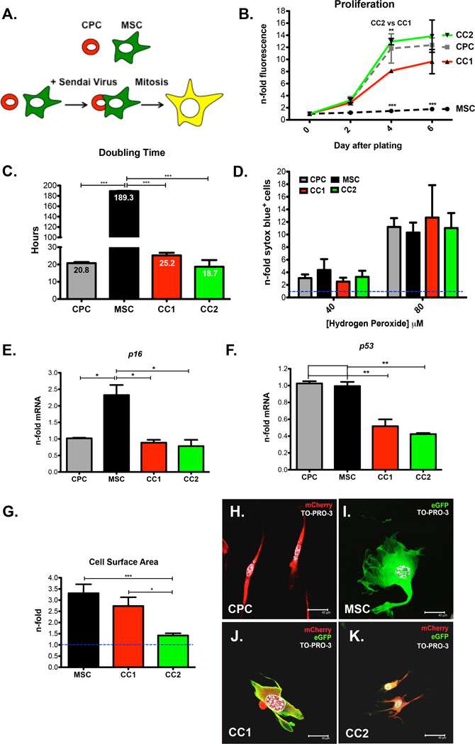
Objective: To demonstrate that CardioChimeras (CCs) formed by fusion between CPCs and MSCs have enhanced reparative potential in a mouse model of myocardial infarction relative to individual stem cells or combined cell delivery.

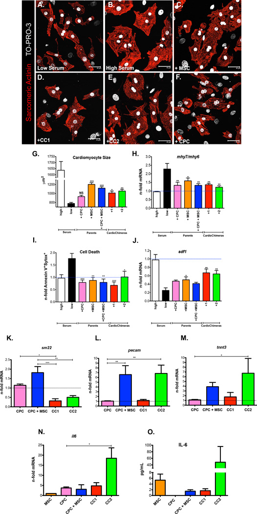
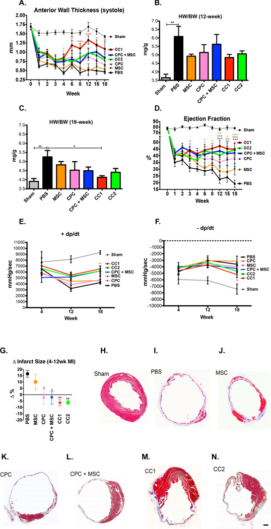
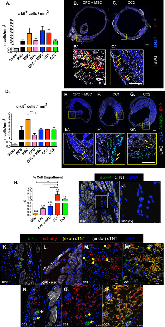
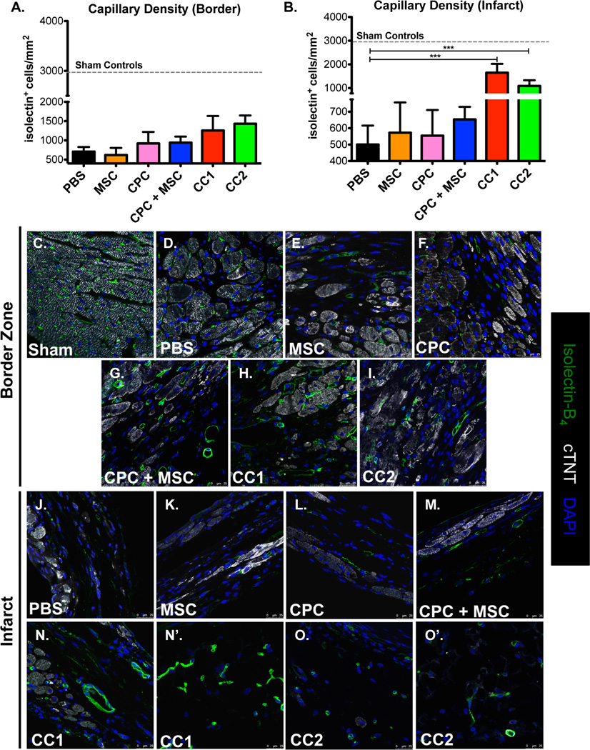
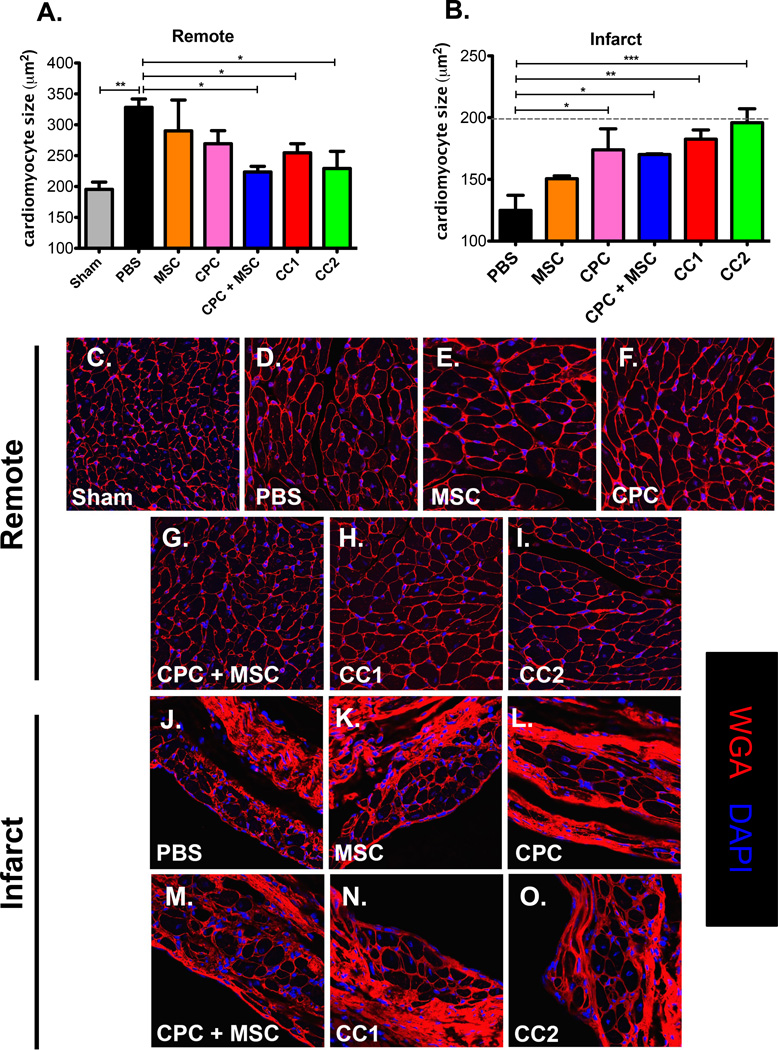
Methods and results: Two distinct and clonally derived CCs, CC1 and CC2, were used for this study. CCs improved left ventricular anterior wall thickness at 4 weeks post injury, but only CC1 treatment preserved anterior wall thickness at 18 weeks. Ejection fraction was enhanced at 6 weeks in CCs, and functional improvements were maintained in CCs and CPC+MSC groups at 18 weeks. Infarct size was decreased in CCs, whereas CPC+MSC and CPC parent groups remained unchanged at 12 weeks. CCs exhibited increased persistence, engraftment, and expression of early commitment markers within the border zone relative to combinatorial and individual cell population-injected groups. CCs increased capillary density and preserved cardiomyocyte size in the infarcted regions suggesting CCs role in protective paracrine secretion.

Conclusions: CCs merge the application of distinct cells into a single entity for cellular therapeutic intervention in the progression of heart failure. CCs are a novel cell therapy that improves on combinatorial cell approaches to support myocardial regeneration.

Keywords: cell fusion; mesenchymal stromal cells; myocardial infarction; myocytes, cardiac; stem cells.


Figures

Figure 1
Figure 1. Phenotypic Characterization of CardioChimeras
(A) Schematic representation of the creation of CardioChimeras. (B) Proliferation of CCs, CPCs and MSCs represented as a fold change relative to day of plating. (C) Cell doubling time in hours. (D) Cell death assay of CCs and parents cells after treatment with 40µM or 80µM hydrogen peroxide represented as a fold change relative to cells not treated with hydrogen peroxide. (E) p16 and (F) p53 gene expression normalized to ribosomal 18s and represented as a fold change relative to CPCs. (G) Cell surface area represented as a fold change normalized to CPCs (blue dashed line, 1.0). Fluorescent images of (H) CPCs, (I) MSCs, (J) CC1 or (K) CC2. * p<0.05, ** p<0.01, *** p<0.001. Scale bar is 40µm.
Figure 2
Figure 2. CardioChimeras promote cell growth and have increased commitment and paracrine gene expression after in vitro co-culture with cardiac myocytes
(A) NRCMs in low serum. (B) NRCMS in high serum. (C) NRCMs in low serum and after the addition of MSCs, (D) CC1, (E) CC2 or (F) CPCs for 24 hours. Cardiac myocytes were visualized by staining with sarcomeric actinin. TO-PRO-3 iodide was used to visualize nuclei. (G) Quantitation of cardiomyocyte size. (H) Gene expression of mhy7 over mhy6 represented as a fold change relative to high serum. (I) Cardiomyocyte cell death. Values are represented as fold change of Annexin V+ and Sytox Blue+ cells relative to high serum. (J) sdf-1 gene expression in cardiac myocytes alone after the addition of stem cells. (K–M) Gene expression in stem cells after a 7-day co-culture with NRCMs. (K) sm22 (L) pecam gene expression. (M and N) il6 gene expression analyzed in stem cells after a 24-hour co-culture with NRCMs. (O) IL-6 expression confirmed by ELISA.(G–J) Statistical values were determined by one-way ANOVA compared to low serum controls. * p<0.05, ** p<0.01, *** p<0.001. Scale bar is 40µm.
Figure 3
Figure 3. CardioChimeras improve left ventricular wall structure and cardiac function after myocardial injury
(A) Longitudinal assessment of anterior wall thickness during systole (mm) over 18 weeks. (B) Heart weight to body weight ratio (mg/g) at 12 WPI (C) 18 WPI. Sample sizes of 3–5 mice per group. (D) Longitudinal assessment of ejection fraction (%). (E) Positive and (F) Negative developed pressure over time represented as mmHg/sec at 4, 12 and 18 WPI. (G) Change in infarct size between 4 and 12 weeks time points. P values were determined by one-way ANOVA compared to PBS treated controls. (H–N) Masson’s Trichrome staining and representative images of infarct size and fibrosis in (H) Sham, (I) PBS, (J) MSC, (K) CPC, (L) CPC + MSC, (M) CC1 and (N) CC2. Sample sizes are specified in the Online Table II. All statistical values were determined by two-way ANOVA compared to PBS treated hearts. * p<0.05, ** p<0.01, *** p<0.001. Colors of asterisk(s) correspond to heart group. Scale bar is 250µm.
Figure 4
Figure 4. CardioChimeras have increased engraftment, expression of cardiomyogenic markers and support the increased presence of c-kit+ cells in the myocardium 12 weeks after damage
(A) Number of c-kit+ cells over the area of left ventricular free wall (mm2) in a 4-week damaged heart. Representative whole heart scans of (B) CPC + MSC and (C) CC2 treated hearts to visualize c-kit+ cells (red). Scale bar is 100µm. (B’) and (C’) C-kit+ cells are identified by yellow arrows. Scale bar is 50µm. (D) Number of c-kit+ cells in 12-week damaged heart.. Representative whole heart scans of (E) CPC + MSC, (F) CC1 and (G) CC2 treated hearts to visualize exogenous mcherry+ cells (green) and c-kit+ cells (red). Scale bar is 100µm. (E’), (F’) and (G’) C-kit+ cells are identified by yellow arrows. Scale bar is 100µm. (H) Cell engraftment efficiency (%). (I) MSC detected by GFP fluorescence at 12 weeks. (J) 2× zoom of a MSC in the border zone area. (K) C-kit+ /mcherry+ CPCs in the border zone area. (L) Mcherry+ CPC in CPC + MSC treated heart. (M) Mcherry+ CC1 visualized in the infarcted area surrounded by c-kit+ cells (green). (M’) Overlay of cTNT (exogenous-cTNT, yellow) in CC1 mcherry labeled cells. (N) Mcherry+ CC2 visualized in the infarcted area surrounded by c-kit+ cells (green). (O) Mcherry+ CC2 (red) visualized in the infarcted area surrounded by c-kit+ cells (green). (O) Overlay of cTNT (exogenous-cTNT, yellow) in CC2 mcherry labeled cells. Endogenous-cTNT (white) labels existing cardiac myocytes. Sample size of 3 mice per group. * p<0.05, ** p<0.01, *** p<0.001.
Figure 5
Figure 5. CardioChimeras increase capillary density in the infarct area
(A) Capillary density in the border zone and (B) Infarcted heart regions. Sample sizes are 3–4 mice per group. Sham controls (dashed line) are represented as control for baseline density of isolectin+ structures per mm2. (C–I) Representative border zone images to visualize isolectin+ structures. (J–O) Representative infarct zone images to visualize and quantitate isolectin+ structures. Green= Isolectin B4, White=cardiac troponin T and Blue= DAPI to stain for nuclei. Scale bar is 25µm.* p<0.05, ** p<0.01, *** p<0.001.
Figure 6
Figure 6. CPC, MSC and CardioChimera treatment antagonizes cardiomyocyte hypertrophy in the remote region and preserves cardiomyocyte size in the infarcted regions
(A) Mean cardiomyocyte size in the remote and (B) Infarct regions. Sample size is 3–4 mice per group. (C–I) Representative images of remote area cardiomyocyte size. (J–O) Representative images of infarct area cardiac myocytes. Red=Wheat germ agglutinin, White=cardiac troponin T and Blue=DAPI to stain for nuclei. Scale bar is 25µm.* p<0.05, ** p<0.01, *** p<0.001.

Comment in

References

    1. Hong KU, Guo Y, Li QH, Cao P, Al-Maqtari T, Vajravelu BN, Du J, Book MJ, Zhu X, Nong Y, Bhatnagar A, Bolli R. C-kit+ cardiac stem cells alleviate post-myocardial infarction left ventricular dysfunction despite poor engraftment and negligible retention in the recipient heart. PLoS One. 2014;9:e96725. - PMC - PubMed
    1. Fischer KM, Cottage CT, Wu W, Din S, Gude NA, Avitabile D, Quijada P, Collins BL, Fransioli J, Sussman MA. Enhancement of myocardial regeneration through genetic engineering of cardiac progenitor cells expressing pim-1 kinase. Circulation. 2009;120:2077–2087. - PMC - PubMed
    1. Mohsin S, Khan M, Toko H, Bailey B, Cottage CT, Wallach K, Nag D, Lee A, Siddiqi S, Lan F, Fischer KM, Gude N, Quijada P, Avitabile D, Truffa S, Collins B, Dembitsky W, Wu JC, Sussman MA. Human cardiac progenitor cells engineered with pim-i kinase enhance myocardial repair. Journal of the American College of Cardiology. 2012;60:1278–1287. - PMC - PubMed
    1. Leri A, Kajstura J, Anversa P. Cardiac stem cells and mechanisms of myocardial regeneration. Physiological reviews. 2005;85:1373–1416. - PubMed
    1. van Berlo JH, Kanisicak O, Maillet M, Vagnozzi RJ, Karch J, Lin SC, Middleton RC, Marban E, Molkentin JD. C-kit+ cells minimally contribute cardiac myocytes to the heart. Nature. 2014;509:337–341. - PMC - PubMed

Publication types

MeSH terms