. 2015 Jul 31:5:12519.
doi: 10.1038/srep12519.

The relationship between nociceptive brain activity, spinal reflex withdrawal and behaviour in newborn infants

Affiliations

The relationship between nociceptive brain activity, spinal reflex withdrawal and behaviour in newborn infants

Caroline Hartley et al. Sci Rep. .

Abstract

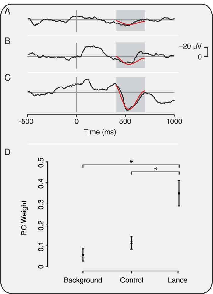
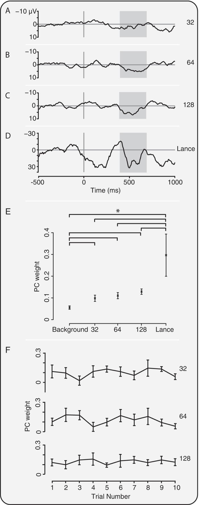
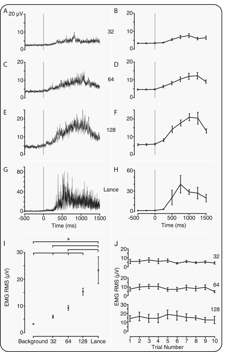
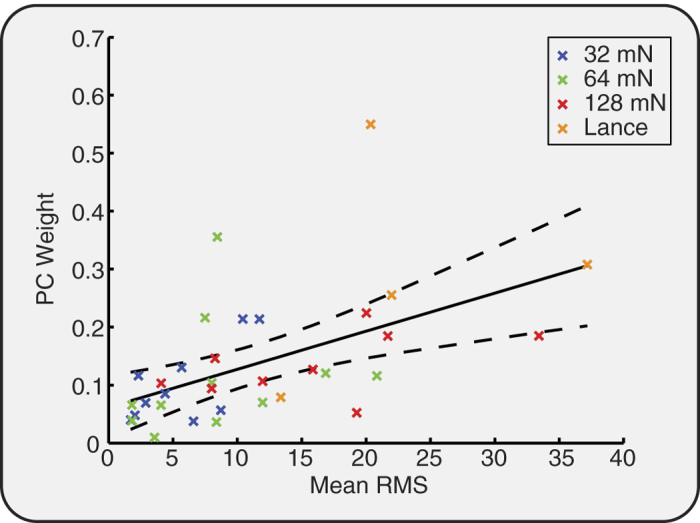
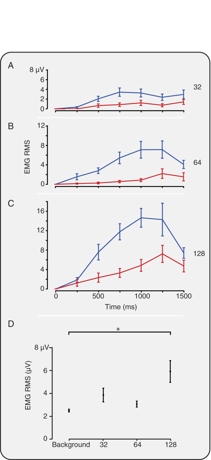
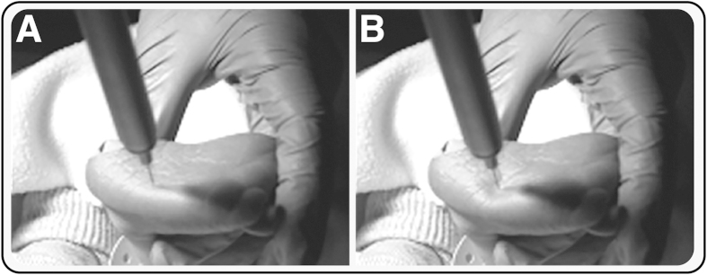
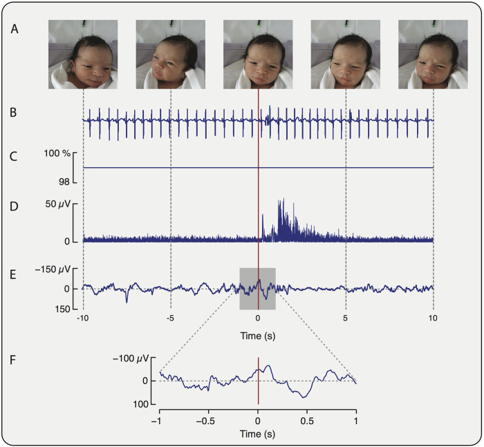
Measuring infant pain is complicated by their inability to describe the experience. While nociceptive brain activity, reflex withdrawal and facial grimacing have been characterised, the relationship between these activity patterns has not been examined. As cortical and spinally mediated activity is developmentally regulated, it cannot be assumed that they are predictive of one another in the immature nervous system. Here, using a new experimental paradigm, we characterise the nociceptive-specific brain activity, spinal reflex withdrawal and behavioural activity following graded intensity noxious stimulation and clinical heel lancing in 30 term infants. We show that nociceptive-specific brain activity and nociceptive reflex withdrawal are graded with stimulus intensity (p < 0.001), significantly correlated (r = 0.53, p = 0.001) and elicited at an intensity that does not evoke changes in clinical pain scores (p = 0.55). The strong correlation between reflex withdrawal and nociceptive brain activity suggests that movement of the limb away from a noxious stimulus is a sensitive indication of nociceptive brain activity in term infants. This could underpin the development of new clinical pain assessment measures.


Figures

Figure 1
Figure 1. Characterisation of nociceptive-specific brain activity.
Average EEG activity recorded during (A) background, (B) in response to non-noxious control stimulation, and (C) following noxious heel lance in 6 infants. The nociceptive-specific principal component (PC) is overlaid in red. The PC weights were significantly greater (*p < 0.05) in response to lance than background and control (D). Error bars indicate standard error of the mean.
Figure 2
Figure 2. Experimental noxious stimuli evoke nociceptive-specific brain activity.
Average traces (without Woody filtering jitter) in response to the (A) 32 mN, (B) 64 mN, and (C) 128 mN experimental noxious stimulus, and (D) clinically required heel lance. (E) PC weight of the nociceptive-specific brain activity was graded with intensity and was significantly different to background data following all the experimental noxious stimuli and heel lance (*p < 0.05). (F) PC weight did not significantly change with stimulus number for any of the experimental noxious stimuli. Error bars indicate standard error of the mean. Grey boxes indicate the time window 400–700 ms.
Figure 3
Figure 3. Experimental noxious stimuli evoke significant reflex withdrawal.
Average EMG response (A,C,E,G) and average RMS (B,D,F,H) to the (A,B) 32 mN, (C,D) 64 mN, and (E,F) 128 mN experimental noxious stimulus and (G,H) clinically required heel lance. (I) The reflex withdrawal was graded with stimulus intensity and for all stimuli the response was significantly different to background activity (*p < 0.05). (J) The reflex withdrawal response was not significantly different across stimulus number for any of the experimental noxious stimuli. Error bars indicate standard error of the mean.
Figure 4
Figure 4. Reflex withdrawal and nociceptive-specific evoked brain activity is correlated.
PC weight of the nociceptive-specific brain activity plotted against average EMG RMS for each infant and each stimulus. The regression line (solid) and 95% confidence intervals (dashed) are shown in black.
Figure 5
Figure 5. Bilateral leg withdrawal occurs with higher intensity noxious stimuli.
EMG RMS in response to the (A) 32 mN, (B) 64 mN, and (C) 128 mN experimental noxious stimulus for the ipsilateral, i.e. stimulated, (blue) and contralateral (red) leg. (D) The average EMG RMS in the contralateral limb in response to stimuli (in the subset of trials when significant ipsilateral reflex withdrawal occurred) and in background activity. Only following the 128 mN stimuli was the response significantly different to background activity (*p < 0.05). Error bars indicate standard error of the mean.
Figure 6
Figure 6. Time-locking of experimental noxious stimuli.
Experimental noxious stimuli were time-locked to EEG and EMG recordings using a high-speed video camera. Example images of (A) the point of first contact of the stimulus with the skin, and (B) the point at which the barrel of the stimulus was first depressed (i.e. the point at which the force was first applied), which was taken as the point of stimulation. We would like to acknowledge Ravi Poorun for taking the photographs and for preparing the images.
Figure 7
Figure 7. Behavioural, physiological, spinal cord and brain activity recorded in response to an experimental noxious stimulus.
Recordings of (A) facial expression; (B) heart rate; (C) oxygen saturation; (D) EMG activity recorded from the biceps femoris of the stimulated leg and (E,F) EEG activity at the Cz electrode site are shown during application of a 128 mN stimulus. (The stimuli was applied at time = 0 seconds, and the facial expression screen shots are shown at −10, −5, 0, 5 and 10 seconds).

References

    1. Craig K. D., Whitfield M. F., Grunau R. V., Linton J. & Hadjistavropoulos H. D. Pain in the preterm neonate: behavioural and physiological indices. Pain 52, 287–299 (1993). - PubMed
    1. Grunau R. V. & Craig K. D. Pain expression in neonates: facial action and cry. Pain 28, 395–410 (1987). - PubMed
    1. Worley A., Fabrizi L., Boyd S. & Slater R. Multi-modal pain measurements in infants. J Neurosci Methods 205, 252–257, 10.1016/j.jneumeth.2012.01.009 (2012). - DOI - PMC - PubMed
    1. Anand K. J. & International Evidence-Based Group for Neonatal Pain. Consensus statement for the prevention and management of pain in the newborn. Arch Pediatr Adolesc Med. 155, 173–180 (2001). - PubMed
    1. Morison S. J., Grunau R. E., Oberlander T. F. & Whitfield M. F. Relations between behavioral and cardiac autonomic reactivity to acute pain in preterm neonates. Clin J Pain. 17, 350–358 (2001). - PMC - PubMed

Publication types