. 2015 Sep 15;309(6):G491-9.
doi: 10.1152/ajpgi.00140.2015. Epub 2015 Jul 30.

Suppression of fibrogenic signaling in hepatic stellate cells by Twist1-dependent microRNA-214 expression: Role of exosomes in horizontal transfer of Twist1

Affiliations

Suppression of fibrogenic signaling in hepatic stellate cells by Twist1-dependent microRNA-214 expression: Role of exosomes in horizontal transfer of Twist1

Li Chen et al. Am J Physiol Gastrointest Liver Physiol. .

Abstract

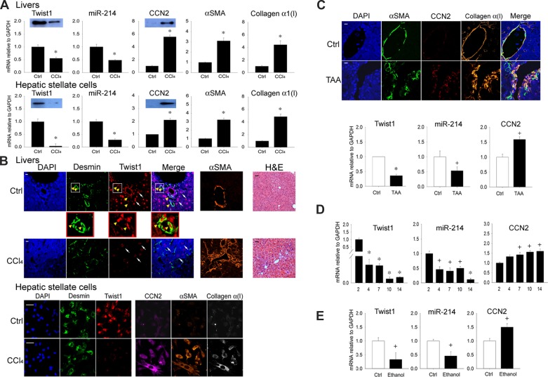
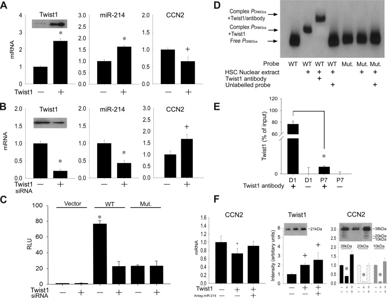
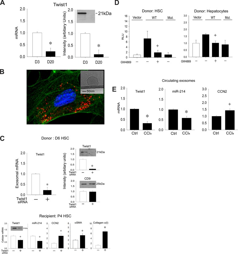
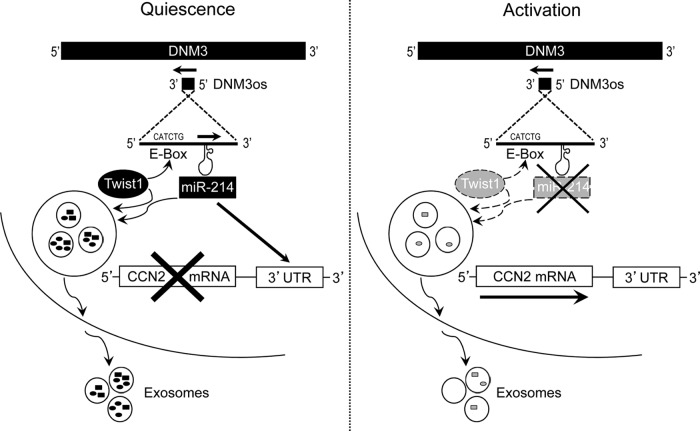
A hallmark of liver fibrosis is the activation of hepatic stellate cells (HSC), which results in their production of fibrotic molecules, a process that is largely regulated by connective tissue growth factor (CCN2). CCN2 is increasingly expressed during HSC activation because of diminished expression of microRNA-214 (miR-214), a product of dynamin 3 opposite strand (DNM3os) that directly suppresses CCN2 mRNA. We show that an E-box in the miR-214 promoter binds the basic helix-loop-helix transcription factor, Twist1, which drives miR-214 expression and results in CCN2 suppression. Twist1 expression was suppressed in HSC of fibrotic livers or in cultured HSC undergoing activation in vitro or after treatment with ethanol. Furthermore, Twist1 decreasingly interacted with DNM3os as HSC underwent activation in vitro. Nanovesicular exosomes secreted by quiescent but not activated HSC contained high levels of Twist1, thus reflecting the suppression of cellular Twist1 during HSC activation. Exosomal Twist1 was intercellularly shuttled between HSC and stimulated expression of miR-214 in the recipient cells, causing expression of CCN2 and its downstream effectors to be suppressed. Additionally, the miR-214 E-box in HSC was also regulated by hepatocyte-derived exosomes, showing that functional transfer of exosomal Twist1 occurs between different cell types. Finally, the levels of Twist1, miR-214, or CCN2 in circulating exosomes from fibrotic mice reflected fibrosis-induced changes in the liver itself, highlighting the potential utility of these and other constituents in serum exosomes as novel circulating biomarkers for liver fibrosis. These findings reveal a unique function for cellular or exosomal Twist1 in CCN2-dependent fibrogenesis.

Keywords: CCN2; E-box; connective tissue growth factor; exosome; fibrosis.


Figures

Fig. 1.
Fig. 1.
Twist1 is expressed at high levels in normal liver or hepatic stellate cells (HSC) and is suppressed during fibrosis. A: expression of Twist1, connective tissue growth factor (CCN2), α-smooth muscle actin (α-SMA), or collagen α1(I) mRNA or microRNA-214 (miR-214) assessed by qRT-PCR and normalized to GAPDH mRNA after administration of oil or CCl4 for 5 wk determined for RNA from either liver tissue (top) or HSC isolated from the livers and maintained in culture for 24 h (bottom) (n = 5 independent experiments performed in triplicate, *P < 0.001 vs. oil control). Insets: detection of Twist1 or CCN2 by Western blotting of 20 μg total protein (β-actin was detected equally in each set of paired samples; data not shown). B, top: hematoxylin and eosin (H&E) staining or immunohistochemical detection of desmin, Twist1, or α-SMA in the livers of mice treated with oil or CCl4 for 5 wk. Immunostained specimens were also stained with 4′,6-diamidino-2-phenylindole (DAPI) nuclear stain (blue). In control livers, Twist1 staining was associated with the desmin-positive quiescent HSC population, with some staining also in hepatocytes. Fibrosis in response to CCl4 was accompanied by HSC activation, as shown by increased α-SMA staining in HSC and a reduction of Twist1 staining in both HSC and hepatocytes. The magnified parts of the figure illustrate Twist1 staining in desmin-positive HSC from normal liver. Bottom: immunocytochemical staining for desmin, α-SMA, Twist1, CCN2, or collagen α1 in HSC from control or CCl4-treated mice obtained as described in A (bars: 20 μm for immunostained sections; 50 μm for H&E-stained sections). C: immunohistochemical detection of α-SMA, CCN2, or collagen α1 (top: bar = 20 μm) or expression of Twist1, miR-214, or CCN2 (bottom) in the livers of mice treated with water (control) or thioacetic acid (TAA) for 5 wk to induce HSC activation and fibrosis (n = 5 independent experiments performed in triplicate, *P < 0.001, +P < 0.05 vs. no treatment). D: Twist1, miR-214, or CCN2 expression analyzed by qRT-PCR and normalized to GAPDH mRNA in primary HSC isolated from livers of normal mice and cultured for up to 14 days (n = 3 independent experiments performed in triplicate, *P < 0.001 vs. day 2, +P < 0.05 vs. day 2). E: qRT-PCR of Twist1, CCN2 mRNA, or miR-214, normalized to GAPDH mRNA, after day 3 primary mouse HSC were incubated in 1% serum for 24 h before 48-h treatment with 0 or 50 mM ethanol (n = 3 independent experiments performed in triplicate, +P < 0.05 vs. control).
Fig. 2.
Fig. 2.
Twist1 regulates CCN2 production via transcriptional control of miR-214 E-box promoter element. Expression of Twist1, CCN2 mRNA, or miR-214 was assessed by qRT-PCR and normalized to GAPDH mRNA in passage 6 (P6) mouse HSC transfected with Twist1 (A) or day 6 primary mouse HSC transfected with 100 nM Twist1 siRNA (B) (n = 3 independent experiments performed in triplicate, *P < 0.001 vs. scramble, +P < 0.05 vs. scramble). Insets: detection of Twist1 by Western blotting of 20 μg total protein (staining for β-actin confirmed equal protein loading; data not shown). C: day 6 primary mouse HSC were transfected with parental pGL 4.11[Luc2P]-vector (Vector), pGL 4.11[Luc2P] containing wild-type mouse dynamin 3 opposite strand (DNM3os) promoter (WT), or a substitution mutation in the DNM3os promoter targeting the E-box (Mut.). After 36 h, firefly luciferase activity in cell lysates was measured and normalized to that of Renilla luciferase (n = 3 independent experiments performed in triplicate, *P < 0.001 vs. vector group). D: nuclear extracts from D1 mHSC underwent EMSA by incubation with oligonucleotide probes corresponding to the wild-type or mutated Twist1 binding site in the DNM3os promoter. Shifted complexes are indicative of binding interactions with the probe; the involvement of Twist1 is indicated by a supershifted complex using Twist1 antibody. Data are representative of 3 independent experiments. E: chromatin immunoprecipitation-PCR of DNM3os DNA in immune complexes generated using Twist1 antibody (+) to pull down endogenous Twist1-DNM3os complexes from day 1 or P7 HSC. Control reactions were performed with normal IgG (-), *P < 0.001. F: qRT-PCR (left) of CCN2 mRNA normalized to GAPDH mRNA, after LX-2 cells were transfected with Twist1 alone or together with miR-214 antagomir (n = 3 independent experiments performed in triplicate, +P < 0.05 vs. nontransfection). Western blot images and quantification thereof (middle and right) show that Twist1 transfection of LX-2 cells caused higher Twist1 but lower CCN2 protein levels but that CCN2 levels were restored to normal levels when Twist1-transfected cells were also transfected with a miR-214 antagomir. The CCN2 Western blot shows the principal CCN2 38-kDa protein and its 10–20-kDa proteolytic cleavage products.
Fig. 3.
Fig. 3.
Identification and intercellular transfer of exosomal Twist1. A: Twist1 mRNA assessed by qRT-PCR and normalized to let-7a (left) or protein assessed by Western blot (right, 5 μg total protein) in exosomes isolated from day 3 or day 20 mouse HSC (n = 3 independent experiments performed in triplicate, *P < 0.001 vs. day 3). B: P6 HSC were incubated for 12 h with exosomes purified from P6 HSC and subsequently stained with PKH-26. Cells were visualized for exosome fluorescence (red) and α-SMA immunofluorescence (green) by confocal microscopy. Inset: appearance of HSC-derived exosomes by cryogenic transmission electron microscopy. C: reduced Twist1 mRNA expression (top, left) or protein levels (top, right) in exosomes produced over 24 h after Twist1 siRNA transfection of day 6 donor HSC and the effect of these exosomes on Twist1, miR-214, CCN2, α-SMA, or collagen α(I) expression assessed by RT-PCR or Twist1 protein levels assessed by Western blot (20 μg total protein, for which β-actin signals were detected equally between samples; data not shown) after being added to recipient P4 HSC for 12 h (bottom). *P < 0.001 and +P < 0.05 vs. control exosomes from nontransfected cells. D: day 6 primary mouse HSC were transfected with parental pGL 4.11[Luc2P]-vector or pGL 4.11[Luc2P] containing WT or mutant mouse DNM3os promoter (see Fig. 2C) and cocultured for 24 h with D1 HSC (left) or AML12 mouse hepatocytes (right) that had been pretreated with or without GW4869 for 24 h. Firefly luciferase activity in cell lysates was measured and normalized to that of Renilla luciferase (n = 3 independent experiments performed in triplicate, +P < 0.05 vs. Vector). E: PureExo exosome isolation kit was used to isolate circulating exosomes from the serum of mice after 5-wk administration of oil or CCl4 Twist1, miR-214, or CCN2 expression in circulating exosomes analyzed by qRT-PCR and normalized to let-7a (n = 5 independent experiments performed in triplicate, *P < 0.001, +P < 0.05 vs. oil group).
Fig. 4.
Fig. 4.
The Twist1-miR-214-CCN2 axis in HSC. In quiescent HSC, Twist1 is expressed at high levels and drives miR-214 expression through its binding of the E-box in DNM3os. One of the targets of miR-214 is the 3′-untranslated region (UTR) of CCN2, resulting in the inhibition of CCN2 in quiescent cells. Twist1 (these results) or miR-214 (4) are exported from quiescent HSC (or hepatocytes) (see Fig. 3D) in exosomes, allowing them to exert epigenetic effects on their targets (miR-214 or CCN2, respectively) after being shuttled to neighboring HSC, causing fibrogenic signaling to be suppressed. HSC activation is characterized by suppressed Twist1 production (the mediators of which have yet to be determined) and reduced activation of the E-box (dashed arrow). In turn, expression of miR-214 and its binding to the CCN2 3′-UTR are reduced, allowing CCN2 expression and its downstream fibrogenic cascades to be manifested. Twist1 or miR-214 is incorporated into exosomes at lower levels (dashed arrows) with the result that exosomes from activated HSC are less effective at suppressing the miR-214-CCN2 axis in neighboring HSC.

References

    1. Bourgeois P, Bolcato-Bellemin AL, Danse JM, Bloch-Zupan A, Yoshiba K, Stoetzel C, Perrin-Schmitt F. The variable expressivity and incomplete penetrance of the twist-null heterozygous mouse phenotype resemble those of human Saethre-Chotzen syndrome. Hum Mol Genet 7: 945–957, 1998. - PubMed
    1. Chairoungdua A, Smith DL, Pochard P, Hull M, Caplan MJ. Exosome release of beta-catenin: a novel mechanism that antagonizes Wnt signaling. J Cell Biol 190: 1079–1091, 2010. - PMC - PubMed
    1. Charrier A, Chen R, Chen L, Kemper S, Hattori T, Takigawa M, Brigstock DR. Exosomes mediate intercellular transfer of pro-fibrogenic connective tissue growth factor (CCN2) between hepatic stellate cells, the principal fibrotic cells in the liver. Surgery 156: 548–555, 2014. - PMC - PubMed
    1. Chen L, Charrier A, Zhou Y, Chen R, Yu B, Agarwal K, Tsukamoto H, Lee LJ, Paulaitis ME, Brigstock DR. Epigenetic regulation of connective tissue growth factor by MicroRNA-214 delivery in exosomes from mouse or human hepatic stellate cells. Hepatology 59: 1118–1129, 2014. - PMC - PubMed
    1. Chen L, Charrier AL, Leask A, French SW, Brigstock DR. Ethanol-stimulated differentiated functions of human or mouse hepatic stellate cells are mediated by connective tissue growth factor. J Hepatol 55: 399–406, 2011. - PMC - PubMed

Publication types

MeSH terms