. 2015 Sep 18;290(38):23064-76.
doi: 10.1074/jbc.M115.648642. Epub 2015 Jul 30.

Regulation of Ergothioneine Biosynthesis and Its Effect on Mycobacterium tuberculosis Growth and Infectivity

Affiliations

Regulation of Ergothioneine Biosynthesis and Its Effect on Mycobacterium tuberculosis Growth and Infectivity

Melissa Richard-Greenblatt et al. J Biol Chem. .

Abstract

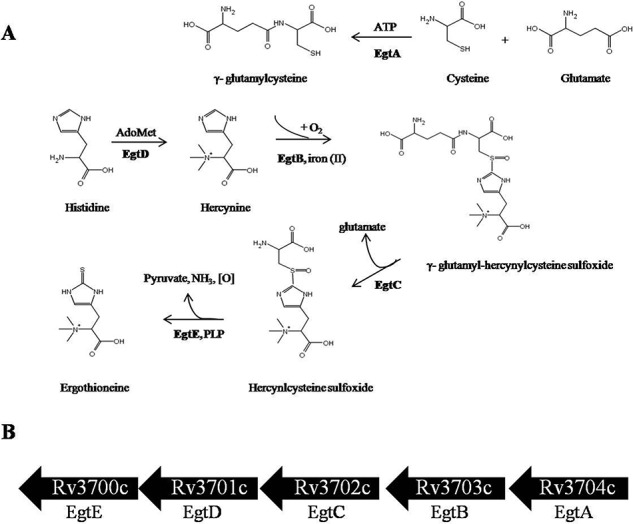
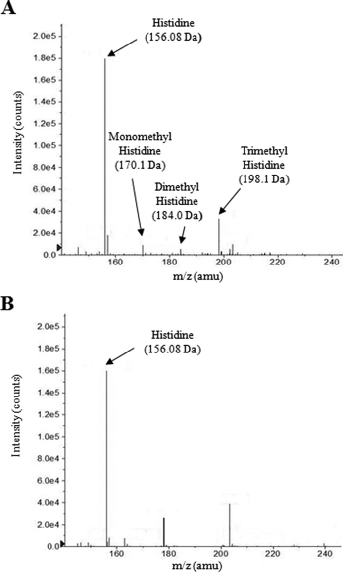
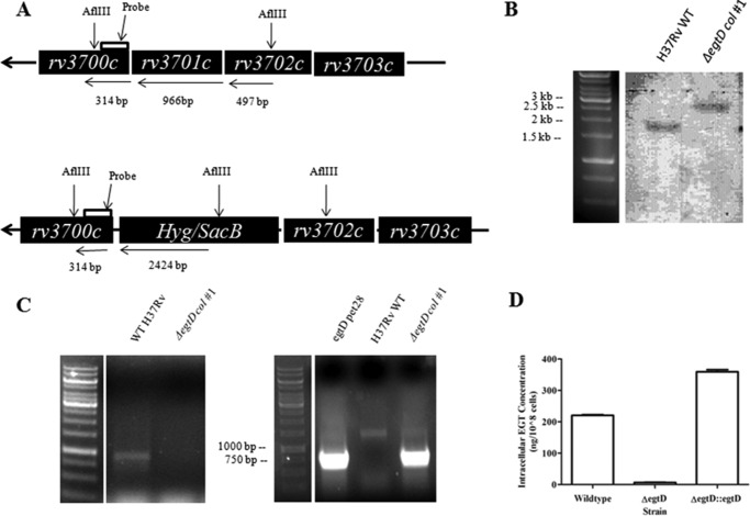
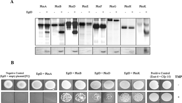
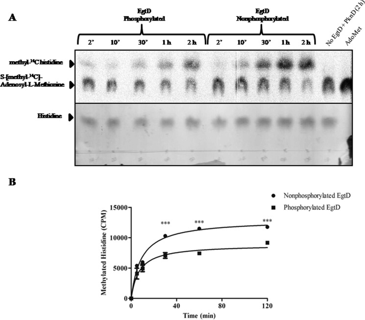
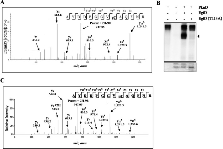
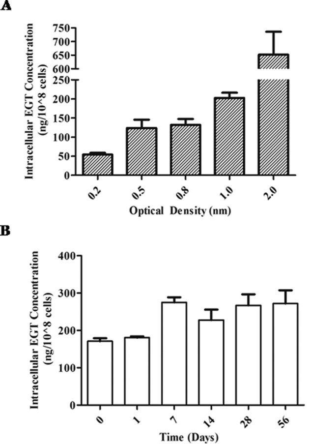
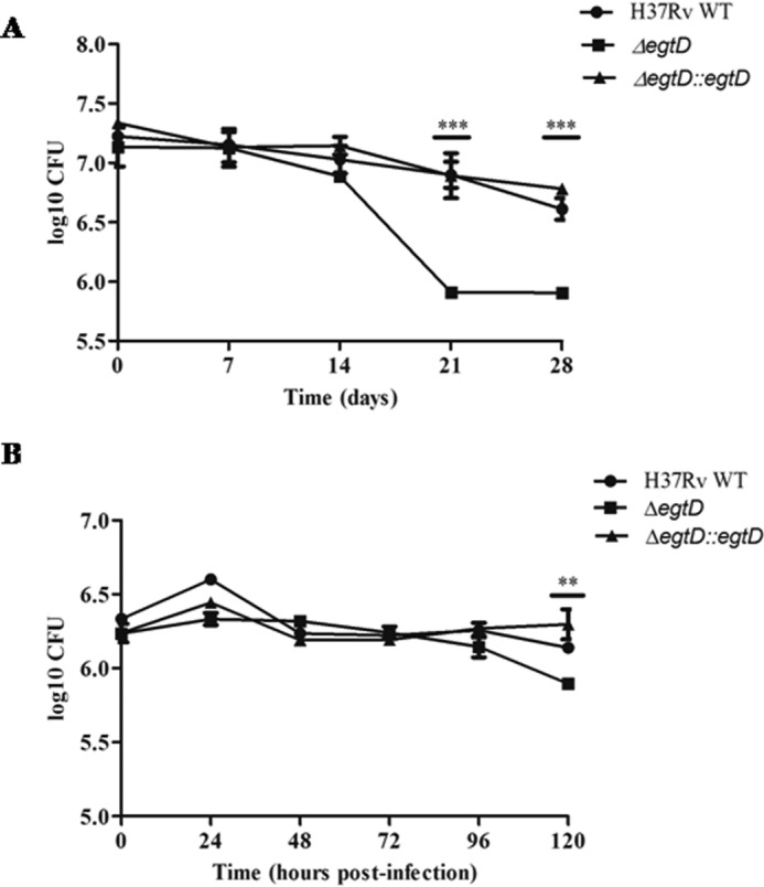
Ergothioneine (EGT) is synthesized in mycobacteria, but limited knowledge exists regarding its synthesis, physiological role, and regulation. We have identified Rv3701c from Mycobacterium tuberculosis to encode for EgtD, a required histidine methyltransferase that catalyzes first biosynthesis step in EGT biosynthesis. EgtD was found to be phosphorylated by the serine/threonine protein kinase PknD. PknD phosphorylates EgtD both in vitro and in a cell-based system on Thr(213). The phosphomimetic (T213E) but not the phosphoablative (T213A) mutant of EgtD failed to restore EGT synthesis in a ΔegtD mutant. The findings together with observed elevated levels of EGT in a pknD transposon mutant during in vitro growth suggests that EgtD phosphorylation by PknD negatively regulates EGT biosynthesis. We further showed that EGT is required in a nutrient-starved model of persistence and is needed for long term infection of murine macrophages.

Keywords: Mycobacterium tuberculosis; bacterial protein kinase; bacterial signal transduction; ergothioneine; histidine methylation; microbiology; thiol.


Figures

FIGURE 1.
FIGURE 1.
EGT biosynthetic pathway in M. tuberculosis. A, EGT biosynthesis occurs through five enzymatic steps, and the genes encode for a γ-glutamylcysteine synthase (EgtA), a formylglycine-generating enzyme-like protein (EgtB), a glutamine amidotransferase (EgtC), a methyltransferase (EgtD), and a pyridoxal 5-phosphate protein (EgtE). The pathway proceeds from l-histidine through the intermediary precursor hercynine, hercynyl γ-glutamylcysteine sulfoxide, and hercynylcysteine sulfoxide. EGT acquires its sulfur from γ-glutamylcysteine. B, in silico analysis identified the gene cluster Rv3700c–Rv3704c to encode for EGT biosynthesis in M. tuberculosis. PLP, pyridoxal 5-phosphate.
FIGURE 2.
FIGURE 2.
Rv3701c encodes for a histidine methyltransferase. The methylation activity of Rv3701c in the presence of AdoMet and histidine was analyzed by ESI-MS. A, Rv3701c catalyzes the methylation of the α-amino nitrogen atom of histidine to form mono-, di-, and trimethylated histidine. B, reaction in the absence of Rv3701c. No methylated histidine products were observed. amu, atomic mass units.
FIGURE 3.
FIGURE 3.
Construction and in vitro characterization of ΔegtD in M. tuberculosis. A, schematic diagram of the Rv3701c region of the chromosome of M. tuberculosis. Genomic DNA was digested with AflIII, and the blot was probed with a digoxigenin-11-dUTP-labeled DNA fragment containing 314 bp of the egtD 3′-flanking sequence. B, confirmation of the ΔegtD mutant through Southern blotting. AflIII-digested genomic DNA gave rise to the expected 1.77-kbp fragment in wild-type M. tuberculosis (lane 1) and 2.74-kbp fragment in the hygromycin-resistant transductant in which egtD was disrupted with the hyg marker (lane 2). C, PCR analysis of the hygromycin-resistant transductant genomic DNA for the ΔegtD. Left panel, PCR amplification of egtD (966 bp). Right panel, PCR amplification of the hygromycin-resistant cassette (∼700 bp), which replaced egtD in the mutants. D, intracellular EGT levels extracted from wild-type M. tuberculosis, ΔegtD M. tuberculosis, and ΔegtD transformed with pMV261:egtD and quantified by ESI LC-MS/MS. Error bars indicate the means ± S.D. of three independent experiments. Col, colony.
FIGURE 4.
FIGURE 4.
EgtD is a substrate of multiple M. tuberculosis STPKs. A, in vitro phosphorylation of EgtD by multiple kinases. M. tuberculosis STPKs purified as GST or His fusions were incubated with His-tagged EgtD and [γ-32P]ATP. Samples were separated by SDS-PAGE and stained with Coomassie Blue followed by visualization by autoradiography. Upper bands represent autophosphorylation activity of each kinase (Pkn); lower bands reflect phosphorylated EgtD. B, interaction between EgtD and M. tuberculosis STPKs facilitates the reassembly of complementary fragments F1 and F2 and fragment F3 of murine dihydrofolate reductase and thus confers M. smegmatis resistance to trimethoprim (TMP). Growth was monitored over 4 days on kanamycin/hygromycin plates supplemented with 0 and 10 μg/ml trimethoprim. Control plates without trimethoprim revealed growth of all strains. Positive Control, M. tuberculosis ESAT-6 (F1 and F2) and CFP-10 (F3); Negative Control, EgtD (F1 and F2) with F3 alone. Experiments are shown in duplicates.
FIGURE 5.
FIGURE 5.
EgtD methylation activity is negatively regulated by phosphorylation. A, phosphorylated and non-phosphorylated EgtD was obtained from an in vitro kinase assay and added to a methylation assay containing S-[methyl-14C]adenosyl-l-methionine (2 μCi/ml). The transfer of methyl-14C to histidine was monitored over a 2-h period and analyzed by one-dimensional TLC using butanol/acetic acid/water (60:15:25, v/v). Upper, detection of [methyl-14C]histidine was performed by autoradiography, exposing the TLC plate to x-ray cassettes for 1 week. Lower, TLC plate developed with ninhydrin to visualize histidine. B, graphical representation of the effect of phosphorylation on EgtD methylation activity from A. ***, p < 0.0005 for comparison of phosphorylated versus non-phosphorylated EgtD. Error bars indicate the means ± S.E. of three independent experiments. ′, minutes.
FIGURE 6.
FIGURE 6.
Identification of EgtD phosphorylation by PknD. A, MS/MS spectra at +2 representing peptide positions 205–218 with a monoisotopic mass of 1,510.69 Da from EgtD phosphorylated by PknD in vitro. Phosphorylation at Thr213 was shown by the “y” C-terminal daughter ion series where all y ions identified lose phosphoric acid (−98 Da) after the phosphorylated residue. pT, phosphothreonine; amu, atomic mass units. B, in vitro kinase assay confirmed Thr213 as the major phosphorylation site of EgtD by PknD. EgtD T213A is defective in phosphorylation. Upper, phosphorimage; lower, Coomassie Blue stain. The arrowhead points to EgtD. C, MS/MS spectra m/z 795.83 (+2) representing peptide positions 205–218 from EgtD phosphorylated in a cell-based system with PknD showing phosphorylation of Thr213. Phosphorylation at Thr213 is shown by the y C-terminal daughter ion series where all y ions after Thr213 lose phosphoric acid.
FIGURE 7.
FIGURE 7.
Phosphorylation of EgtD reduces EGT levels in M. tuberculosis. A, electrocompetent H37Rv M. tuberculosis cells were transformed with pMV261_egtD_WT, pMV261_egtD_T213A, and pMV261_egtD_T213E to allow for the constitutive expression of the egtD alleles under the control of the hsp60 promoter. Bacteria were harvested at mid-log phase, washed with purified water, and lysed in 70% acetonitrile. Bacterial lysates were collected and analyzed for EGT by ESI LC-MS. EGT intracellular levels were normalized to the number of cells. B, CDC1551 wild type and a PknD:Tn (point of insertion at bp 1166) generously provided by The John Hopkins Mutant Library were grown to mid-log phase prior to extraction, and EGT was quantified by ESI LC-MS. The results presented for both A and B are expressed as the mean of three independent experiments ±S.E. (error bars). **, p < 0.005; ***, p < 0.0005.
FIGURE 8.
FIGURE 8.
Intracellular EGT levels in H37Rv wild type under nutrient rich and starvation conditions. A, ESI LC-MS/MS quantification of intracellular EGT levels of M. tuberculosis at different stages of growth. Cultures were grown in Middlebrook 7H9 supplemented with 0.2% glycerol, 10% OADC, and 0.05% tyloxapol, and EGT was extracted from each culture at various optical densities. B, monitoring intracellular EGT levels under starved cultures. M. tuberculosis was starved in standing cultures for up to 6 weeks in PBS containing 0.05% tyloxapol. EGT was extracted from M. tuberculosis at weekly intervals for quantification by ESI LC-MS/MS. Both A and B are expressed as mean of three independent experiments ± S.E. (error bars).
FIGURE 9.
FIGURE 9.
Role of EgtD in the viability of M. tuberculosis under nutrient starvation and during macrophage infection. A, survival of H37Rv WT, ΔegtD, and ΔegtD::egtD strains in 4-week-starved cultures. M. tuberculosis was incubated as standing cultures at 37 °C and starved in PBS with 0.05% tyloxapol. Samples were taken on a weekly basis to assess viability by cfu counts. The results of the three experiments (A–C) are representative of two independent experiments ±S.E. (error bars). ***, p < 0.0001. D, replication and survival of H37Rv WT, ΔegtD, and the corresponding complemented strain (ΔegtD::egtD) in J774A.1 macrophages infected at a multiplicity of infection of 5:1. cfu were calculated at the specified time points. Results are representative of two independent experiments ±S.E. (error bars). **, p < 0.01.

Similar articles

Cited by

References

    1. Kumar A., Farhana A., Guidry L., Saini V., Hondalus M., Steyn A. J. (2011) Redox homeostasis in mycobacteria: the key to tuberculosis control? Expert Rev. Mol. Med. 13, e39. - PMC - PubMed
    1. Newton G. L., Arnold K., Price M. S., Sherrill C., Delcardayre S. B., Aharonowitz Y., Cohen G., Davies J., Fahey R. C., Davis C. (1996) Distribution of thiols in microorganisms: mycothiol is a major thiol in most actinomycetes. J. Bacteriol. 178, 1990–1995 - PMC - PubMed
    1. Rawat M., Newton G. L., Ko M., Martinez G. J., Fahey R. C., Av-Gay Y. (2002) Mycothiol-deficient Mycobacterium smegmatis mutants are hypersensitive to alkylating agents, free radicals, and antibiotics. Antimicrob. Agents Chemother. 46, 3348–3355 - PMC - PubMed
    1. Ung K. S., Av-Gay Y. (2006) Mycothiol-dependent mycobacterial response to oxidative stress. FEBS Lett. 580, 2712–2716 - PubMed
    1. Rawat M., Uppal M., Newton G., Steffek M., Fahey R. C., Av-Gay Y. (2004) Targeted mutagenesis of the Mycobacterium smegmatis mca gene, encoding a mycothiol-dependent detoxification protein. J. Bacteriol. 186, 6050–6058 - PMC - PubMed

Publication types

LinkOut - more resources