Review
. 2015 Aug 1;26(15):2727-32.
doi: 10.1091/mbc.E14-04-0940.

Assembly of hair bundles, an amazing problem for cell biology

Affiliations
Review

Assembly of hair bundles, an amazing problem for cell biology

Peter-G Barr-Gillespie. Mol Biol Cell. .

Abstract

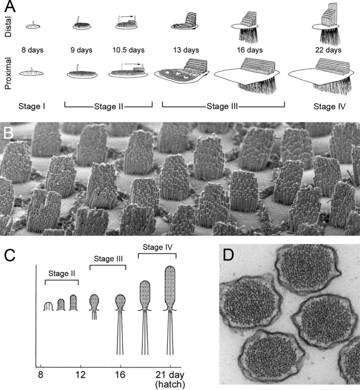
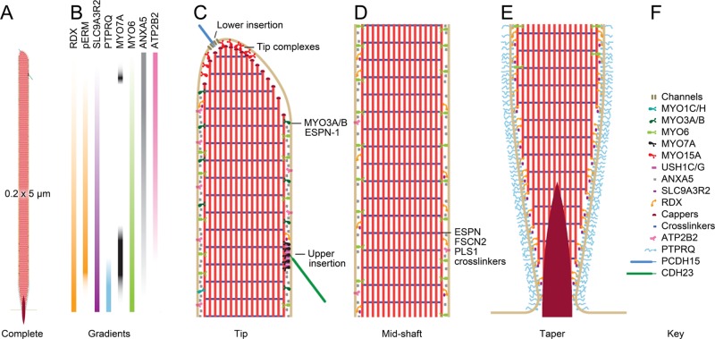
The hair bundle--the sensory organelle of inner-ear hair cells of vertebrates--exemplifies the ability of a cell to assemble complex, elegant structures. Proper construction of the bundle is required for proper mechanotransduction in response to external forces and to transmit information about sound and movement. Bundles contain tightly controlled numbers of actin-filled stereocilia, which are arranged in defined rows of precise heights. Indeed, many deafness mutations that disable hair-cell cytoskeletal proteins also disrupt bundles. Bundle assembly is a tractable problem in molecular and cellular systems biology; the sequence of structural changes in stereocilia is known, and a modest number of proteins may be involved.


Figures

FIGURE 1:
FIGURE 1:
Development of the hair bundle. (A) Progression of hair-bundle morphology in chick cochlea. (B) Chick cochlea hair bundles at hatch. Hair cells imaged are at an intermediate position between the apical and basal extremes. Note the cell-to-cell reproducibility of the numbers of stereocilia, their orientation, and their lengths. Hair bundles are ∼10 μm tall. (C) Progression of stereocilia actin paracrystal development in chick cochlea. (D) Actin paracrystals in mouse vestibular stereocilia. Note the stereocilium-to-stereocilium reproducibility of the size and organization of the actin cores. Stereocilia are ∼200 nm in diameter. A and C are reproduced from Tilney et al. (1992) with permission.
FIGURE 2:
FIGURE 2:
Model chick vestibular stereocilium. Scale model of stereocilium showing selected molecules; these molecules are drawn in at the approximate density for each as determined by mass spectrometry experiments (Shin et al., 2013). (A) Complete stereocilium at low magnification showing dimensions. (B) Selected molecular gradients in the stereocilia. See Shin et al. (2013) for details. (C) High-magnification view of the tip region of the stereocilium. The two insertions of the tip link are highlighted; the lower insertion includes transduction channels, and the upper insertion has USH1C, USH1G, and MYO7A. In addition, the MYO15A tip complex is indicated; DFNB31, EPS8, and perhaps EPS8L2 are part of this complex. MYO3A and MYO3B are found, along with their cargo ESPN-1, in the tip region as well. (D) The stereocilia shaft is made of parallel actin filaments cross-linked by a variety of proteins, most prominently ESPN, FSCN2, and PLS1. Major components of the shaft include the calcium pump ATP2B2, the membrane-associated ANXA5, the actin-to-membrane connector radixin (RDX), and SLC9A3R2, a ligand for RDX. RDX with the activating phosphorylation is found above the taper region. (E) The taper region. Most actin filaments terminate at the membrane, but a few project through into the soma as the rootlet (density). The lipid phosphatase PTPRQ is concentrated in the taper region, as is unactivated RDX. (F) Key to molecules included in the diagram.

References

    1. Avenarius MR, Saylor KW, Lundeberg MR, Wilmarth PA, Shin JB, Spinelli KJ, Pagana JM, Andrade L, Kachar B, Choi D, et al. Correlation of actin crosslinker and capper expression levels with stereocilia growth phases. Mol Cell Proteomics. 2014;13:606–620. - PMC - PubMed
    1. Belyantseva IA, Boger ET, Naz S, Frolenkov GI, Sellers JR, Ahmed ZM, Griffith AJ, Friedman TB. Myosin-XVa is required for tip localization of whirlin and differential elongation of hair-cell stereocilia. Nat Cell Biol. 2005;7:148–156. - PubMed
    1. Beurg M, Fettiplace R, Nam JH, Ricci AJ. Localization of inner hair cell mechanotransducer channels using high-speed calcium imaging. Nat Neurosci. 2009;12:553–558. - PMC - PubMed
    1. Bruckner A, Polge C, Lentze N, Auerbach D, Schlattner U. Yeast two-hybrid, a powerful tool for systems biology. Int J Mol Sci. 2009;10:2763–2788. - PMC - PubMed
    1. Burns JC, On D, Baker W, Collado MS, Corwin JT. Over half the hair cells in the mouse utricle first appear after birth, with significant numbers originating from early postnatal mitotic production in peripheral and striolar growth zones. J Assoc Res Otolaryngol. 2012;13:609–627. - PMC - PubMed

Publication types

LinkOut - more resources