

Melatonin modulates endoplasmic reticulum stress and Akt/GSK3-beta signaling pathway in a rat model of renal warm ischemia reperfusion
- PMID: 26229743
- PMCID: PMC4502281
- DOI: 10.1155/2015/635172
Melatonin modulates endoplasmic reticulum stress and Akt/GSK3-beta signaling pathway in a rat model of renal warm ischemia reperfusion
Abstract
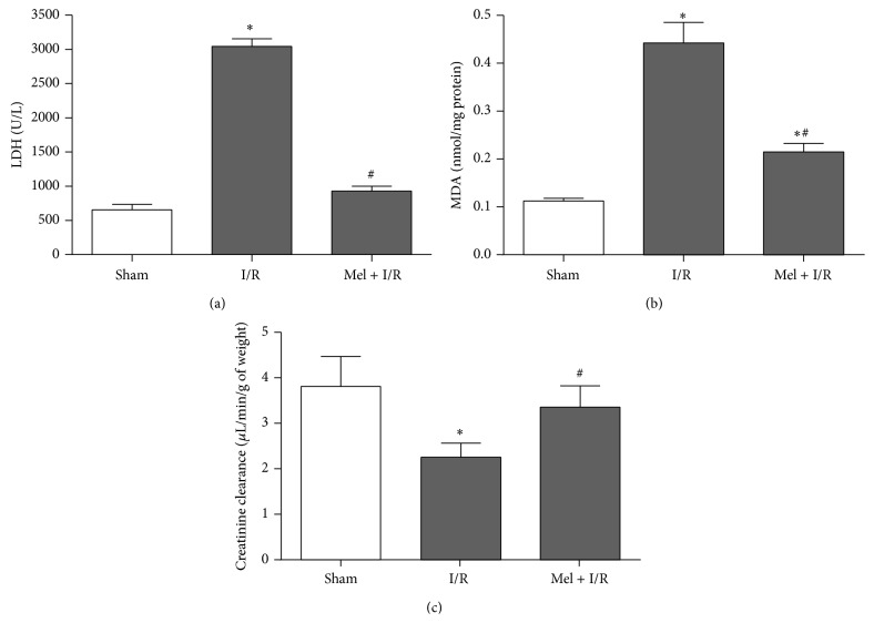
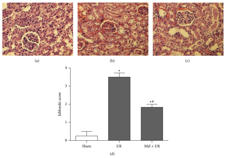
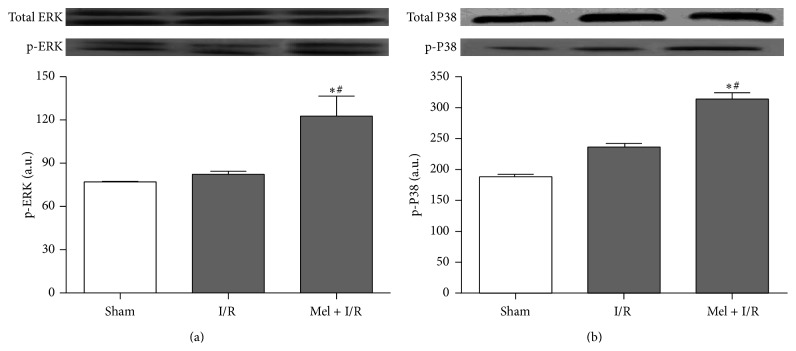
Melatonin (Mel) is widely used to attenuate ischemia/reperfusion (I/R) injury in several organs. Nevertheless, the underlying mechanisms remain unclear. This study was conducted to explore the effect of Mel on endoplasmic reticulum (ER) stress, Akt and MAPK cascades after renal warm I/R. Eighteen Wistar rats were randomized into three groups: Sham, I/R, and Mel + I/R. The ischemia period was 60 min followed by 120 min of reperfusion. Mel (10 mg/kg) was administrated 30 min prior to ischemia. The creatinine clearance, MDA, LDH levels, and histopathological changes were evaluated. In addition, Western blot was performed to study ER stress and its downstream apoptosis as well as phosphorylation of Akt, GSK-3β, VDAC, ERK, and P38. Mel decreased cytolysis and lipid peroxidation and improved renal function and morphology compared to I/R group. Parallely, it significantly reduced the ER stress parameters including GRP 78, p-PERK, XBP 1, ATF 6, CHOP, and JNK. Simultaneously, p-Akt level was significantly enhanced and its target molecules GSK-3β and VDAC were inhibited. Furthermore, the ERK and P38 phosphorylation were evidently augmented after Mel administration in comparison to I/R group. In conclusion, Mel improves the recovery of renal function by decreasing ER stress and stimulating Akt pathway after renal I/R injury.
Figures


Similar articles
-
Attenuation of endoplasmic reticulum stress and mitochondrial injury in kidney with ischemic postconditioning application and trimetazidine treatment.J Biomed Sci. 2012 Aug 1;19(1):71. doi: 10.1186/1423-0127-19-71. J Biomed Sci. 2012. PMID: 22853733 Free PMC article.
-
Melatonin reduces PERK-eIF2α-ATF4-mediated endoplasmic reticulum stress during myocardial ischemia-reperfusion injury: role of RISK and SAFE pathways interaction.Apoptosis. 2016 Jul;21(7):809-24. doi: 10.1007/s10495-016-1246-1. Apoptosis. 2016. PMID: 27170343
-
Thymoquinone prevents endoplasmic reticulum stress and mitochondria-induced apoptosis in a rat model of partial hepatic warm ischemia reperfusion.Biomed Pharmacother. 2017 Oct;94:964-973. doi: 10.1016/j.biopha.2017.08.018. Epub 2017 Aug 12. Biomed Pharmacother. 2017. PMID: 28810534
-
Role of Zinc Supplementation on Ischemia/Reperfusion Injury in Various Organs.Biol Trace Elem Res. 2020 Jul;196(1):1-9. doi: 10.1007/s12011-019-01892-3. Epub 2019 Dec 11. Biol Trace Elem Res. 2020. PMID: 31828721 Review.
-
Protein kinases in organ ischemia and reperfusion.J Invest Surg. 2008 Jul-Aug;21(4):215-26. doi: 10.1080/08941930802130149. J Invest Surg. 2008. PMID: 18615319 Review.
Cited by
-
Melatonin Modulates Neuronal Cell Death Induced by Endoplasmic Reticulum Stress under Insulin Resistance Condition.Nutrients. 2017 Jun 10;9(6):593. doi: 10.3390/nu9060593. Nutrients. 2017. PMID: 28604593 Free PMC article.
-
Melatonin attenuates acute kidney ischemia/reperfusion injury in diabetic rats by activation of the SIRT1/Nrf2/HO-1 signaling pathway.Biosci Rep. 2019 Jan 15;39(1):BSR20181614. doi: 10.1042/BSR20181614. Print 2019 Jan 31. Biosci Rep. 2019. PMID: 30578379 Free PMC article.
-
CD47 blockade reduces ischemia/reperfusion injury in donation after cardiac death rat kidney transplantation.Am J Transplant. 2018 Apr;18(4):843-854. doi: 10.1111/ajt.14523. Epub 2017 Nov 1. Am J Transplant. 2018. PMID: 28975767 Free PMC article.
-
Mechanism of tauroursodeoxycholic acid-mediated neuronal protection after acute spinal cord injury through AKT signaling pathway in rats.Int J Clin Exp Pathol. 2020 Sep 1;13(9):2218-2227. eCollection 2020. Int J Clin Exp Pathol. 2020. PMID: 33042326 Free PMC article.
-
Glycogen synthesis and beyond, a comprehensive review of GSK3 as a key regulator of metabolic pathways and a therapeutic target for treating metabolic diseases.Med Res Rev. 2022 Mar;42(2):946-982. doi: 10.1002/med.21867. Epub 2021 Nov 3. Med Res Rev. 2022. PMID: 34729791 Free PMC article. Review.
References
-
- Arfian N., Emoto N., Vignon-Zellweger N., Nakayama K., Yagi K., Hirata K.-I. ET-1 deletion from endothelial cells protects the kidney during the extension phase of ischemia/reperfusion injury. Biochemical and Biophysical Research Communications. 2012;425(2):443–449. doi: 10.1016/j.bbrc.2012.07.121. - DOI - PubMed
Publication types
MeSH terms
Substances
LinkOut - more resources
Full Text Sources
Other Literature Sources
Research Materials
Miscellaneous
