. 2014 Oct 27;49(6):555-64.
doi: 10.1016/j.rboe.2013.10.002. eCollection 2014 Nov-Dec.

Osteochondroma: ignore or investigate?

Affiliations

Osteochondroma: ignore or investigate?

Antônio Marcelo Gonçalves de Souza et al. Rev Bras Ortop. .

Abstract

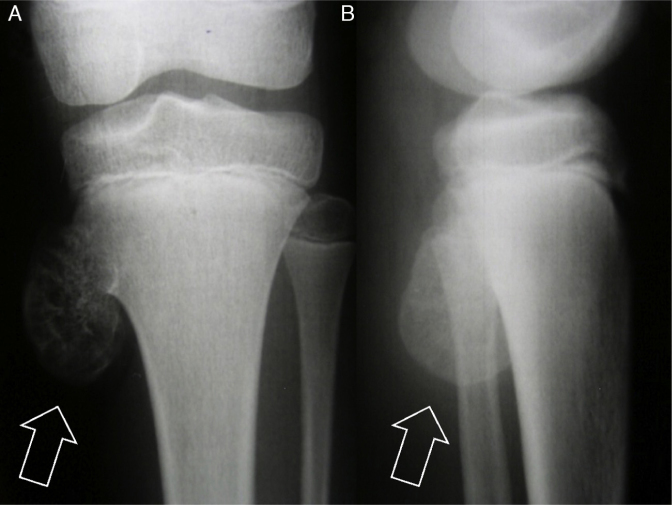
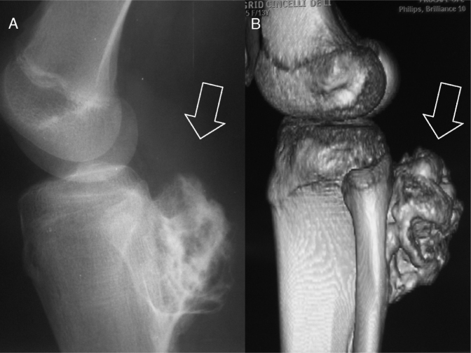
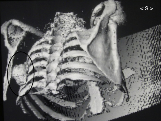
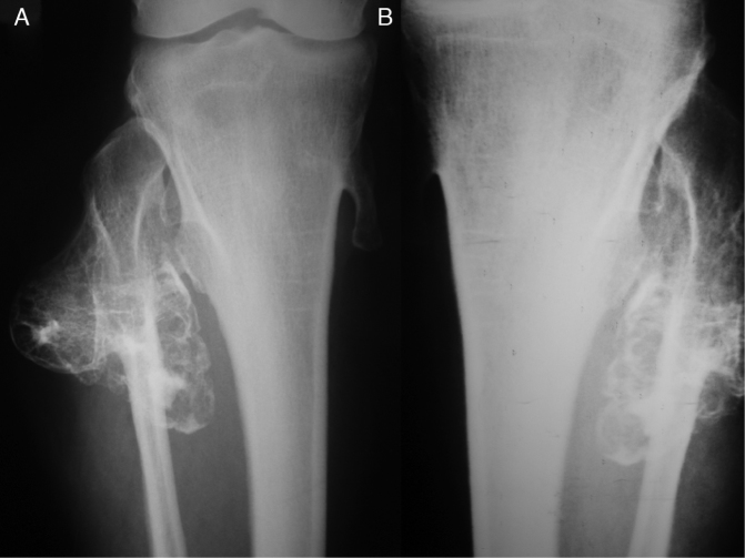
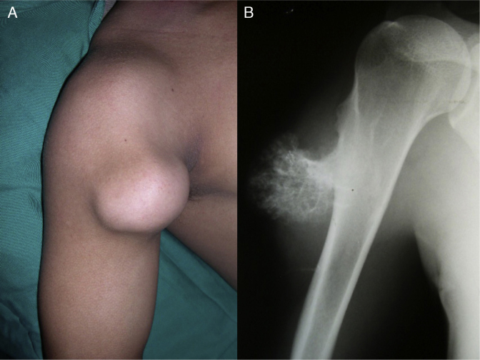
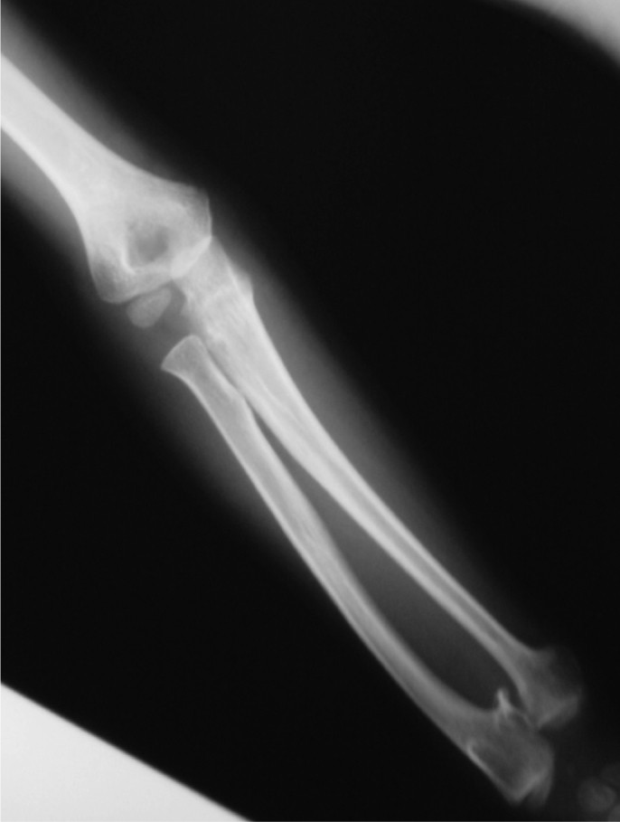
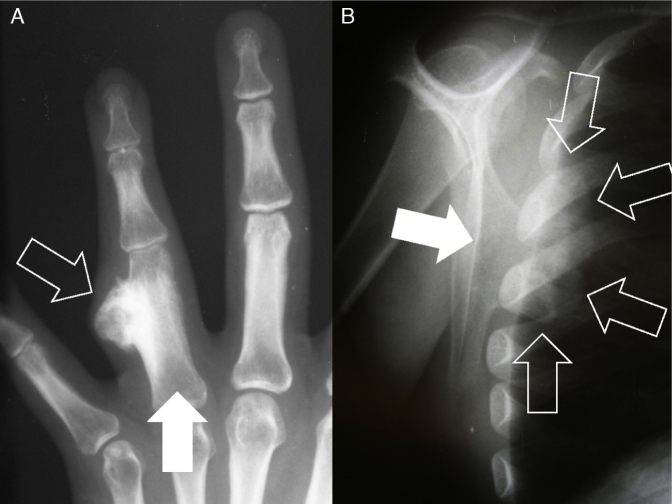
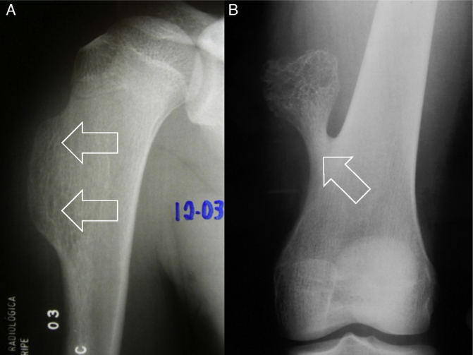
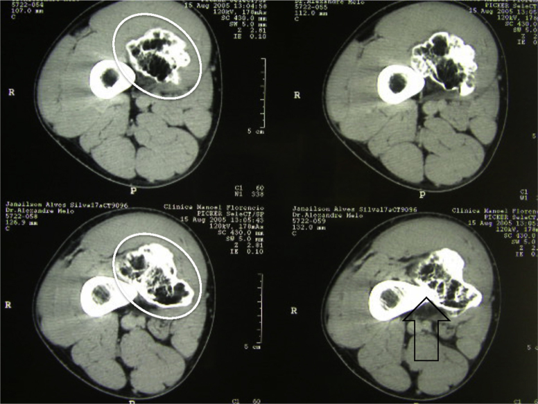
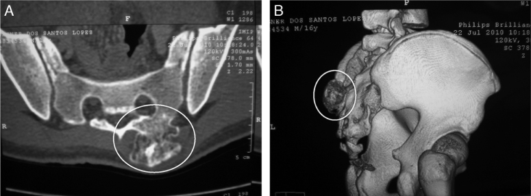
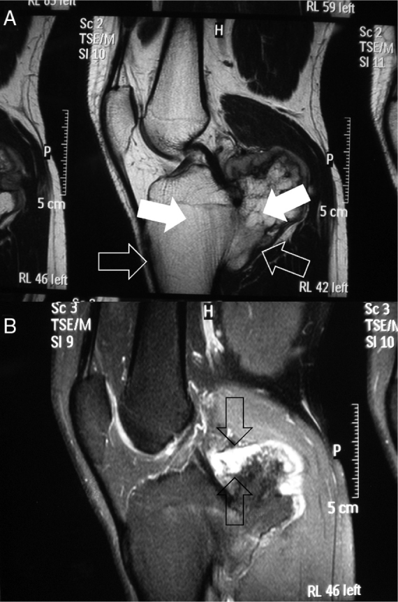
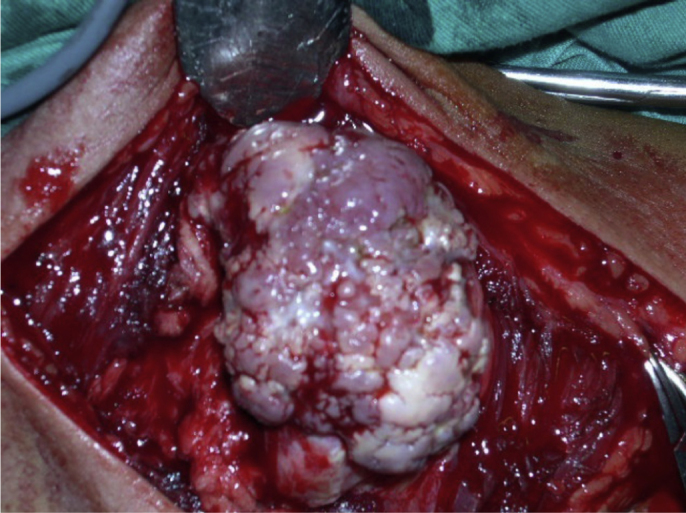
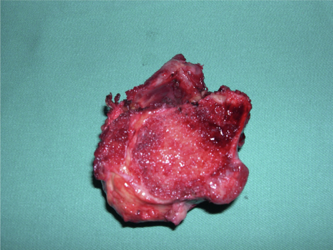
Osteochondromas are bone protuberances surrounded by a cartilage layer. They generally affect the extremities of the long bones in an immature skeleton and deform them. They usually occur singly, but a multiple form of presentation may be found. They have a very characteristic appearance and are easily diagnosed. However, an atypical site (in the axial skeleton) and/or malignant transformation of the lesion may sometimes make it difficult to identify osteochondromas immediately by means of radiographic examination. In these cases, imaging examinations that are more refined are necessary. Although osteochondromas do not directly affect these patients' life expectancy, certain complications may occur, with varying degrees of severity.

Osteocondromas são protuberâncias ósseas envolvidas por uma camada de cartilagem. Atingem, habitualmente, as extremidades dos ossos longos no esqueleto imaturo e os deformam. Em geral são únicos, mas a forma de apresentação múltipla pode ser encontrada. De aspecto bastante característico, são de fácil diagnóstico. Contudo, por vezes, a localização atípica (esqueleto axial) e/ou a malignização da lesão podem dificultar a sua pronta identificação por exames radiográficos. Nesses casos, exames de imagem mais apurados são necessários. Apesar de não afetarem diretamente a expectativa de vida do portador, algumas complicações, com variados graus de gravidade, podem ocorrer.

Keywords: Bone neoplasms; Osteochondroma/diagnosis; Osteochondroma/etiology; Osteochondroma/physiopathology.


Figures

Fig. 1
Fig. 1
Anteroposterior (AP) radiograph (A) and lateral radiograph (B) of the left knee. Note exostosis (osteochondroma – arrows) in the proximal region of the tibia in a skeletally immature patient.
Fig. 2
Fig. 2
The long bones of the lower limbs (knee region) are most commonly affected. (A) Simple lateral radiograph. (B) Computed tomography with 3D reconstruction. Note lesion (arrows) in the proximal region of the tibia.
Fig. 3
Fig. 3
Image of 3D reconstruction from computed tomography of chest. Note single exostosis inside black oval figure in the region of the body of the left scapula, beside the ribs.
Fig. 4
Fig. 4
Hereditary multiple exostosis. (A and B) In the knees, radiographs showing multiple lesions in the proximal regions of the tibias and fibulas.
Fig. 5
Fig. 5
In the clinical examination (A), painless slowly growing bulging of hardened consistency is sometimes observed. (B) Radiograph of the proximal region of the right humerus of the same patient.
Fig. 6
Fig. 6
Radiograph of an individual with hereditary multiple exostosis. Note the deformity of the forearm (due to shortening of the ulna).
Fig. 7
Fig. 7
Radiographs showing projecting osteochondromas (open arrows) in different types of bone. (A) In the long bones (for example, the phalanx – filled arrow), the standard radiographic views (two images in orthogonal planes) are sufficient for the diagnosis. (B) However, in planar bones (for example, the scapula – filled arrow) and irregular bones, exostoses may not be so evident on simple radiographs alone.
Fig. 8
Fig. 8
Different types of osteochondroma. Note that in examination (A), the lesion on the humerus is sessile (with wide base – arrows), while in (B), it is pedicled or pedunculated (narrow base [arrow], i.e. less in relation to its height).
Fig. 9
Fig. 9
Axial computed tomography slices from the distal region of the thigh. Detail from exostosis in the medial region (white oval figure). Note continuity of the lesion with the cortical bone (open black arrow) and its relationship with the adjacent soft tissues.
Fig. 10
Fig. 10
Computed tomography images facilitate locating the exostoses (white oval figures) at anatomical sites of greater complexity (such as the spine–sacral region). (A) Axial image. (B) 3D reconstruction.
Fig. 11
Fig. 11
Magnetic resonance images. (A) T1-weighted sagittal image (note hyposignal of the cortical bone and the lesion [open arrows] and hypersignal of the bone medulla in both [filled arrows]). (B) T2-weighted sagittal image (note that the greatest thickness of the cartilaginous cover was around 1.5 cm [between arrows]).
Fig. 12
Fig. 12
Intraoperative photograph of excision of an osteochondroma. Note its multilobulated surface and cartilage cover.
Fig. 13
Fig. 13
Surgical resection (specimen) was chosen for this exostosis that was causing vascular compression in the popliteal region.

References

    1. Khurana J., Abdul-Karim F., Bovée J.V.M. Osteochondroma. In: Fletcher C.D., Unni K.K., Mertens F., editors. Pathology and genetics of tumours of the soft tissues and bones. IARC Press; Lyon: 2002. pp. 234–237.
    1. Murphey M.D., Choi J.J., Kransdorf M.J., Flemming D.J., Gannon F.H. Imaging of osteochondroma: variants and complications with radiologic–pathologic correlation. Radiographics. 2000;20(5):1407–1434. - PubMed
    1. Costeira O. Farmoquímica; Rio de Janeiro: 2001. Termos e expressões da prática médica.
    1. Unni K.K. 5th ed. Thomas; Springfield: 1996. Osteochondroma. Dahlin's bone tumors: general aspects and data on 11,087 cases; pp. 11–23.
    1. Dorfman H.D., Czerniak B. Mosby; St. Louis: 1998. Osteochondroma. Bone tumors; pp. 331–346.

LinkOut - more resources