

Femoropatellar radiographic alterations in cases of anterior cruciate ligament failure
- PMID: 26229895
- PMCID: PMC4519644
- DOI: 10.1016/j.rboe.2015.01.005
Femoropatellar radiographic alterations in cases of anterior cruciate ligament failure
Abstract
Objective: To make a comparative analysis on three femoropatellar radiographic parameters, between knees with chronic failure of the anterior cruciate ligament (ACL) and normal knees.
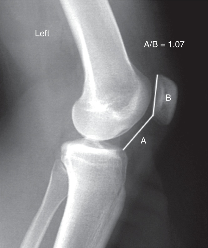
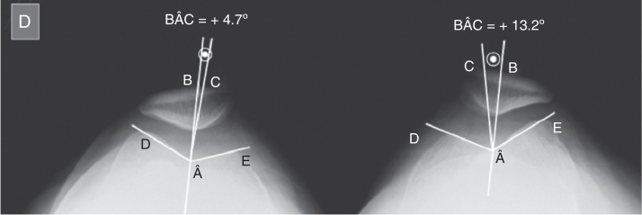
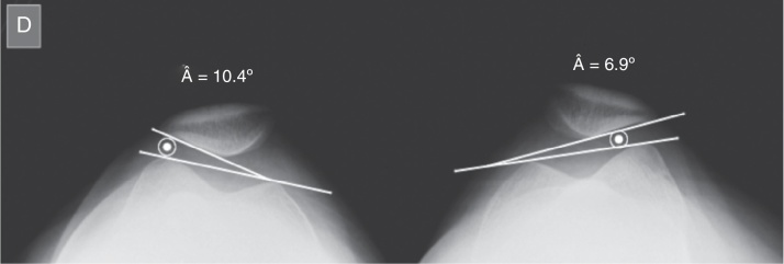
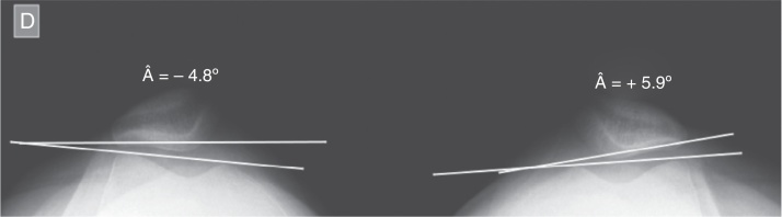
Methods: Thirty volunteer patients with a diagnosis of unilateral isolated chronic ACL injury for more than one year and a normal contralateral knee were selected. Digital radiographs were produced for all the patients, on both knees in absolute lateral view at 30° of flexion, with and without load-bearing on one leg, and in axial view of the patella at 30°. The Caton-Deschamps patellar height index, Merchant patellar congruence angle and Laurin lateral patellar tilt angle were measured on the radiographs obtained from the normal knees and knees with ACL injuries, and comparative analysis was performed between these two groups.
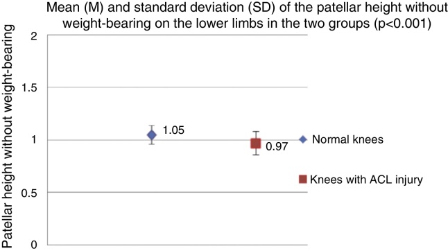
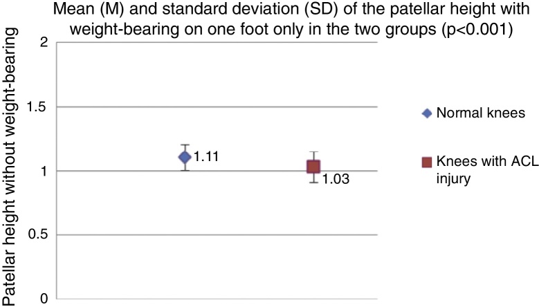
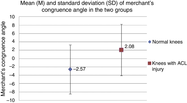
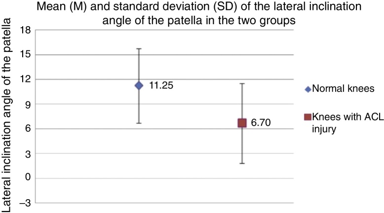
Results: The patellar height was statistically significantly lower (p < 0.001) in the knees with ACL failure than in the normal knees, both on radiographs without loading and on those with single-foot loading. The Merchant patellar congruence angle was significantly smaller (p < 0.001) in the normal knees and the lateral patellar tilt angle was smaller (p < 0.001) in the knees with ACL failure.
Conclusion: Chronic ACL failure gave rise to a statistically significant change in the femoropatellar radiographic values studied (p < 0.001). Knees with injuries to this ligament presented lower patellar height values, greater tilt and lateral displacement of the patella, in relation to the femoral trochlea, in comparison with the normal contralateral knees.
Objetivo: Análise comparativa de três parâmetros radiográficos femoropatelares entre joelhos com insuficiência crônica do ligamento cruzado anterior (LCA) e joelhos normais.
Métodos: Foram selecionados 30 pacientes voluntários com diagnóstico de lesão crônica isolada unilateral do LCA havia mais de um ano e joelho contralateral normal. Todos os pacientes foram submetidos a radiografias digitais de ambos os joelhos nas incidências em perfil absoluto a 30° de flexão, com e sem carga monopodal, e axial de patela a 30°. Foram mensurados, nas radiografias obtidas, o índice de altura patelar de Caton-Deschamps, o ângulo de congruência patelar de Merchant e o ângulo de inclinação lateral da patela, descrito por Laurin, nos joelhos normais e nos joelhos com lesão do LCA e foi feita análise comparativa entre esses dois grupos.
Resultados: A altura patelar foi inferior, de forma estatisticamente significante (p < 0,001), nos joelhos com insuficiência do LCA em comparação com os joelhos normais, tanto nas radiografias sem carga quanto nas com carga monopodal. O ângulo de congruência patelar de Merchant foi significativamente menor (p < 0,001) nos joelhos normais e o ângulo de inclinação lateral da patela foi inferior (p < 0,001) nos joelhos com insuficiência do LCA.
Conclusão: A insuficiência crônica do LCA alterou de forma estatisticamente significante (p < 0,001) os valores dos parâmetros radiográficos femoropatelares estudados. Joelhos com lesão desse ligamento apresentaram menores valores de altura patelar, maior inclinação e deslocamento laterais da patela em relação à tróclea femoral comparados com os joelhos contralaterais normais.
Keywords: Anterior cruciate ligament; Joint instability; Patellofemoral joint.
Figures
References
-
- Ristanis S., Giakas G., Papageorgiou C.D., Moraiti T., Stergiou N., Georgoulis A.D. The effects of anterior cruciate ligament reconstruction on tibial rotation during pivoting after descending stairs. Knee Surg Sports Traumatol Arthrosc. 2003;11(6):360–365. - PubMed
-
- Dennis D.A., Mahfouz M.R., Komistek R.D., Hoff W. In vivo determination of normal and anterior cruciate ligament-deficient knee kinematics. J Biomech. 2005;38(2):241–253. - PubMed
-
- Buckwalter J.A., Lane N.E. Athletics and osteoarthritis. Am J Sports Med. 1997;25(6):873–881. - PubMed
-
- Georgoulis A.D., Papadonikolakis A., Papageorgiou C.D., Mitsou A., Stergiou N. Three-dimensional tibiofemoral kinematics of the anterior cruciate ligament-deficient and reconstructed knee during walking. Am J Sports Med. 2003;31(1):75–79. - PubMed
-
- Andriacchi T.P., Dyrby C.O. Interactions between kinematics and loading during walking for the normal and ACL deficient knee. J Biomech. 2005;38(2):293–298. - PubMed
LinkOut - more resources
Full Text Sources
Other Literature Sources