. 2015 Aug 11;54(31):4927-35.
doi: 10.1021/acs.biochem.5b00335. Epub 2015 Jul 31.

Biochemical and Spectroscopic Studies of Epoxyqueuosine Reductase: A Novel Iron-Sulfur Cluster- and Cobalamin-Containing Protein Involved in the Biosynthesis of Queuosine

Affiliations

Biochemical and Spectroscopic Studies of Epoxyqueuosine Reductase: A Novel Iron-Sulfur Cluster- and Cobalamin-Containing Protein Involved in the Biosynthesis of Queuosine

Zachary D Miles et al. Biochemistry. .

Abstract

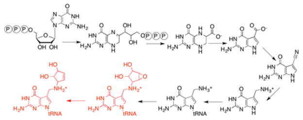
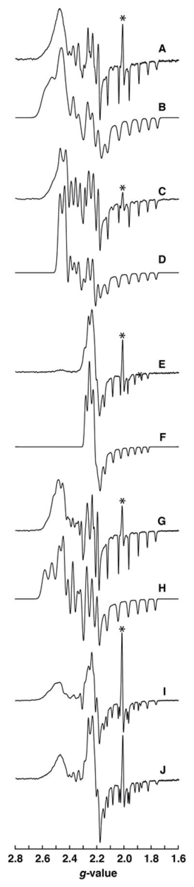
Queuosine is a hypermodified nucleoside present in the wobble position of tRNAs with a 5'-GUN-3' sequence in their anticodon (His, Asp, Asn, and Tyr). The 7-deazapurine core of the base is synthesized de novo in prokaryotes from guanosine 5'-triphosphate in a series of eight sequential enzymatic transformations, the final three occurring on tRNA. Epoxyqueuosine reductase (QueG) catalyzes the final step in the pathway, which entails the two-electron reduction of epoxyqueuosine to form queuosine. Biochemical analyses reveal that this enzyme requires cobalamin and two [4Fe-4S] clusters for catalysis. Spectroscopic studies show that the cobalamin appears to bind in a base-off conformation, whereby the dimethylbenzimidazole moiety of the cofactor is removed from the coordination sphere of the cobalt but not replaced by an imidazole side chain, which is a hallmark of many cobalamin-dependent enzymes. The bioinformatically identified residues are shown to have a role in modulating the primary coordination sphere of cobalamin. These studies provide the first demonstration of the cofactor requirements for QueG.


Figures

Figure 1
Figure 1
Biosynthesis of queuosine.
Figure 2
Figure 2. A representative multiple sequence alignment of QueG homologs and in vivo alanine scanning of conserved residues of QueG
Conserved cysteine residues required for iron-sulfur cluster formation are in red, the conserved DYH motif is shown in orange, and other conserved residues mutated in the in vivo alanine scanning experiment are highlighted in blue. The accession numbers for the sequences are as follows: E. coli (EGI43095), Vibrio cholerae (WP_000386321), Pseudomonas putida (WP_014754621), Mesorhizobium loti (WP_010912587), Maricaulis maris (WP_011642061), and Bacillus subtilis (NP_388772).
Figure 3
Figure 3. Purification, UV/visible spectrum, and quaternary state determination of QueG
A. An SDS-PAGE gel of purified QueG. Based on the gel, QueG is >95% pure with a molecular weight consistent with the predicted weight of 48.7 kDa. B. A size exclusion chromatogram of protein standards (shown in dotted black line) superimposed with a chromatogram of protein standards including purified QueG enzyme (shown in solid black line). Based on the elution profiles, only the peak pertaining to the ~44 kDa standard changes with inclusion of QueG in the solution mixture. QueG is ~48.7 kDa leading to the increase in only the absorbance of the concurrent standard peak. The standard peaks (from left to right) are: thyroglobulin (670 kDa), γ-globulin (150 kDa), ovalbumin (44 kDa), myoglobin (17 kDa) and vitamin B12 (1350 Da). C. A UV/visible spectrum of the purified enzyme contains features consistent with inclusion of both iron-sulfur clusters (~420 nm) and cob(II)alamin (~475 nm) as cofactors.
Figure 4
Figure 4. Iron-sulfur clusters and cobalamin are required for QueG activity in vitro
Extracted ion chromatograms of digested nucleosides of oQ (m/z = 426) and Q (m/z = 410) from oQ stem loop used as the substrate in an in vitro activity assay of QueG. Protein was assayed for activity before and after reconstitution in the presence or absence of exogenous hydroxocobalamin. The appearance of turnover in only enzyme with reconstituted iron-sulfur clusters and added hydroxocobalamin demonstrates the necessity of both for activity.
Figure 5
Figure 5. In vivo QueG mutagenesis
The relative abundances of Q and oQ were calculated from the extracted ion chromatograms of oQ (blue) and Q (red) present in total RNA nucleosides from overnight growths of ΔqueG E. coli overexpressing the indicated variant form of QueG (B. subtilis numbering). All values are normalized to the sum of both oQ and Q and given as a percentage of the total.
Figure 6
Figure 6. EPR of wild-type QueG and variants
The EPR spectra and simulations of A. wild-type and variants C. D104A, E. Y105A, G. H106A, and I. D134A of QueG in the cob(II)alamin form. Simulations are shown in B, D, F, H, and J for wild-type and variants in the same order as above. The * denotes signal that results from degradation of the iron-sulfur clusters during purification and reconstitution.

References

    1. Machnicka MA, Milanowska K, Osman Oglou O, Purta E, Kurkowska M, Olchowik A, Januszewski W, Kalinowski S, Dunin-Horkawicz S, Rother KM, Helm M, Bujnicki JM, Grosjean H. MODOMICS: a database of RNA modification pathways--2013 update. Nucleic Acids Res. 2012;41:D262–D267. - PMC - PubMed
    1. Urbonavičius J, Meškys R, Grosjean H. Biosynthesis of wyosine derivatives in tRNA(Phe) of Archaea: role of a remarkable bifunctional tRNA(Phe):m1G/imG2 methyltransferase. RNA. 2014;20:747–753. - PMC - PubMed
    1. Young AP, Bandarian V. Radical mediated ring formation in the biosynthesis of the hypermodified tRNA base wybutosine. Curr Opin Chem Biol. 2013;17:613–618. - PMC - PubMed
    1. El Yacoubi B, Bailly M, de Crécy-Lagard V. Biosynthesis and Function of Posttranscriptional Modifications of Transfer RNAs. Annu Rev Genet. 2012;46:69–95. - PubMed
    1. Jackman JE, Alfonzo JD. Transfer RNA modifications: nature’s combinatorial chemistry playground. WIREs RNA. 2013;4:35–48. - PMC - PubMed

Publication types

MeSH terms