

Recent insights into the transcriptional control of the Tcra/Tcrd locus by distant enhancers during the development of T-lymphocytes
- PMID: 26230488
- PMCID: PMC4802736
- DOI: 10.1080/21541264.2015.1078429
Recent insights into the transcriptional control of the Tcra/Tcrd locus by distant enhancers during the development of T-lymphocytes
Abstract
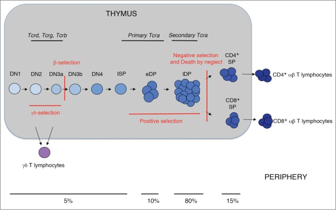
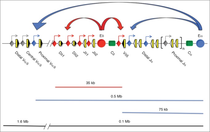
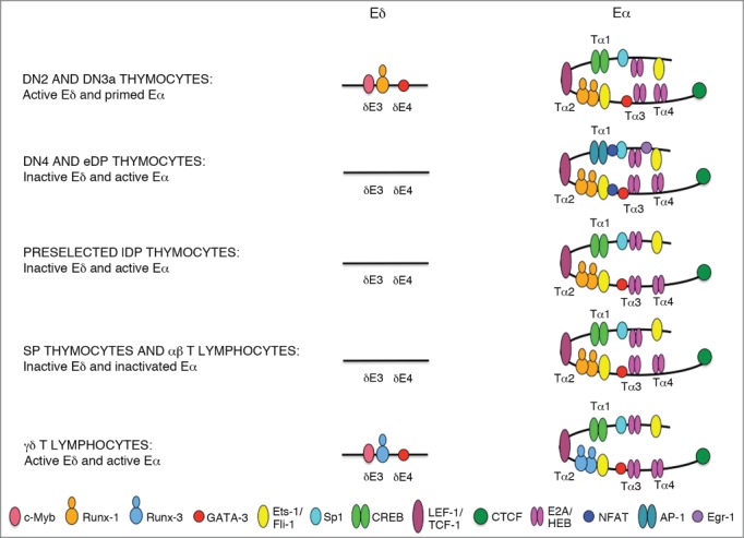
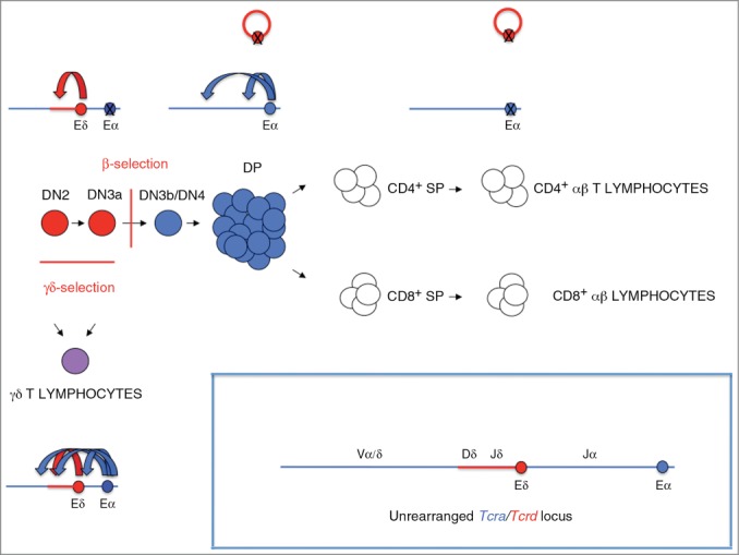
Tcra/Tcrd includes 2 genes with distinct developmental programs controlled by 2 distant enhancers, Eα and Eδ. These enhancers work as a developmental switch during thymocyte development and they are essential for generation of αβ and γδ T-lymphocytes. Tcra and Tcrd transit from an unrearranged configuration to a rearranged configuration during T-cell development. Eα and Eδ are responsible for transcription of their respective unrearranged genes in thymocytes but are dispensable for such functions in the context of the rearranged genes in mature T-cells. Interestingly, Eα activates transcription of the rearranged Tcrd in γδ T-lymphocytes but it is inactive in αβ T-lymphocytes.
Keywords: T-cell development; T-cell receptor; T-lymphocytes; chromatin, enhancer, promoter; thymocytes; transcription.
Figures
Similar articles
-
T-cell receptor α enhancer is inactivated in αβ T lymphocytes.Proc Natl Acad Sci U S A. 2015 Apr 7;112(14):E1744-53. doi: 10.1073/pnas.1406551112. Epub 2015 Mar 23. Proc Natl Acad Sci U S A. 2015. PMID: 25831496 Free PMC article.
-
Tcrd Rearrangement Redirects a Processive Tcra Recombination Program to Expand the Tcra Repertoire.Cell Rep. 2017 Jun 6;19(10):2157-2173. doi: 10.1016/j.celrep.2017.05.045. Cell Rep. 2017. PMID: 28591585 Free PMC article.
-
Trav15-dv6 family Tcrd rearrangements diversify the Tcra repertoire.J Exp Med. 2022 Feb 7;219(2):e20211581. doi: 10.1084/jem.20211581. Epub 2021 Dec 15. J Exp Med. 2022. PMID: 34910107 Free PMC article.
-
Chromatin Dynamics and the Development of the TCRα and TCRδ Repertoires.Adv Immunol. 2015;128:307-61. doi: 10.1016/bs.ai.2015.07.005. Epub 2015 Aug 15. Adv Immunol. 2015. PMID: 26477370 Review.
-
Regulation of T-cell Receptor Gene Expression by Three-Dimensional Locus Conformation and Enhancer Function.Int J Mol Sci. 2020 Nov 11;21(22):8478. doi: 10.3390/ijms21228478. Int J Mol Sci. 2020. PMID: 33187197 Free PMC article. Review.
Cited by
-
Current insights in mouse iNKT and MAIT cell development using single cell transcriptomics data.Semin Immunol. 2022 Mar;60:101658. doi: 10.1016/j.smim.2022.101658. Epub 2022 Sep 28. Semin Immunol. 2022. PMID: 36182863 Free PMC article. Review.
-
The insulator EACBE regulates V(D)J recombination of Tcrd gene by modulating chromatin organization.Front Immunol. 2025 Jul 17;16:1613621. doi: 10.3389/fimmu.2025.1613621. eCollection 2025. Front Immunol. 2025. PMID: 40746544 Free PMC article.
-
A pooled CRISPR screen identifies the Tα2 enhancer element as a driver of TRA expression in a subset of mature human T lymphocytes.Front Immunol. 2025 Mar 14;16:1536003. doi: 10.3389/fimmu.2025.1536003. eCollection 2025. Front Immunol. 2025. PMID: 40160815 Free PMC article.
-
Direct regulation of TCR rearrangement and expression by E proteins during early T cell development.WIREs Mech Dis. 2022 Nov;14(6):e1578. doi: 10.1002/wsbm.1578. Epub 2022 Jul 18. WIREs Mech Dis. 2022. PMID: 35848146 Free PMC article. Review.
-
A BCWD-resistant line of rainbow trout exhibits higher abundance of IgT+ B cells and heavy chain tau transcripts compared to a susceptible line following challenge with Flavobacterium psychrophilum.Dev Comp Immunol. 2017 Sep;74:190-199. doi: 10.1016/j.dci.2017.04.019. Epub 2017 May 4. Dev Comp Immunol. 2017. PMID: 28479345 Free PMC article.
References
-
- Heinz S, Romanoski CE, Benner C, Glass CK. The selection and functions of cell type-specific enhancers. Nat Rev 2015; 16:144-53; PMID:25650801; http://dx.doi.org/10.1038/nrm3949 - DOI - PMC - PubMed
-
- Rothenberg EV, Tanghon T. Molecular genetics of T cell development. Annu Rev Immunol 2005; 23:601-49; PMID:15771582; http://dx.doi.org/10.1146/annurev.immunol.23.021704.115737 - DOI - PubMed
-
- Taghon T, Yui MA, Pant R, Diamond RA, Rothenberg EV. Developmental and molecular characterization of emerging β- and γδ-selected pre-T cells in the adult mouse thymus. Immunity 2006; 24:53-64; PMID:16413923; http://dx.doi.org/10.1016/j.immuni.2005.11.012 - DOI - PubMed
-
- Seitan VC, Hao B, Tachibana-Konwalski K, Lavagnolli T, Mira-Bontenbal H, Brown KE, Teng G, Carroll T, Terry A, Horan K, et al.. A role for cohesin in T-cell-receptor rearrangement and thymocyte differentiation. Nature 2011; 476:467-71; PMID:21832993; http://dx.doi.org/10.1038/nature10312 - DOI - PMC - PubMed
-
- Brekelmans P, van Soest P, Voerman J, Platenburg PP, Leenen PJ, van Ewijk W. Transferrin receptor expression as a marker of immature cycling thymocytes in the mouse. Cell Immunol 1994; 159:331-9; PMID:7994765; http://dx.doi.org/10.1006/cimm.1994.1319 - DOI - PubMed
Publication types
MeSH terms
Substances
LinkOut - more resources
Full Text Sources
Other Literature Sources