. 2015 Jul 31;10(7):e0134523.
doi: 10.1371/journal.pone.0134523. eCollection 2015.

Circulating CCR7+ICOS+ Memory T Follicular Helper Cells in Patients with Multiple Sclerosis

Affiliations

Circulating CCR7+ICOS+ Memory T Follicular Helper Cells in Patients with Multiple Sclerosis

Xueli Fan et al. PLoS One. .

Abstract

Objective: This study is aimed at examining the potential roles of circulating memory T follicular helper (Tfh) cells in patients with multiple sclerosis (MS).

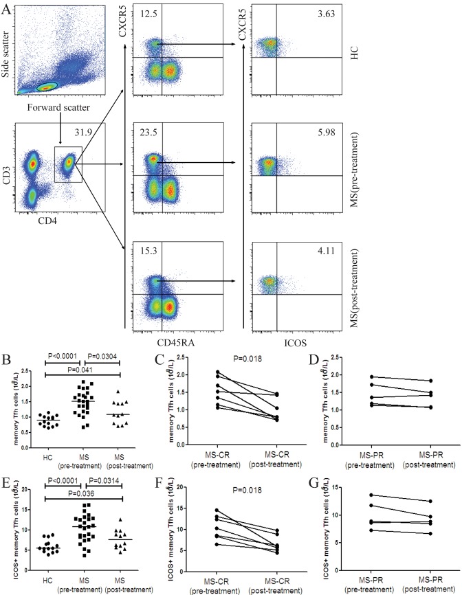
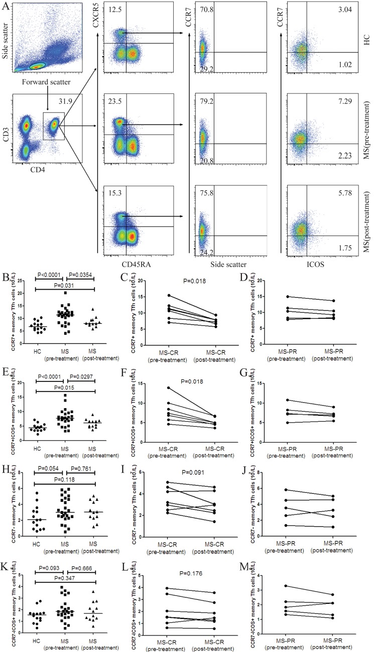
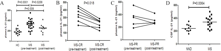
Methods: The numbers of different subsets of circulating memory Tfh cells in 25 patients with relapsed MS before and after treatment as well as 14 healthy controls (HC) were examined by flow cytometry. The levels of plasma IL-21 in all patients and cerebrospinal fluid (CSF) IL-21 in some MS patients and controls with non-inflammatory neuronal diseases (NND) were measured by ELISA.

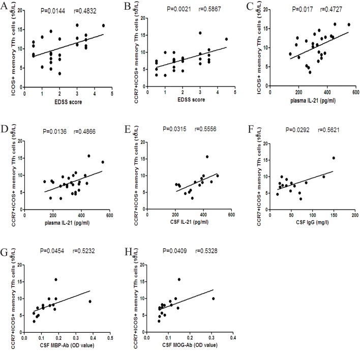
Results: In comparison with that in the HC, the numbers of circulating CD3+CD4+CXCR5+CD45RA-, ICOS+, CCR7+ and CCR7+ICOS+ memory Tfh cells and the levels of plasma IL-21 significantly increased in MS patients, but significantly decreased in the patients with complete remission (CR). The levels of CSF IL-21 were significantly higher in the MS patients than that in the NND patients. The numbers of CCR7+ICOS+ memory Tfh cells were positively correlated with the EDSS scores, the levels of plasma and CSF IL-21, IgG, MBP-Ab or MOG-Ab.

Conclusions: Our findings indicated that circulating memory Tfh cells, especially CCR7+ICOS+ memory Tfh cells, may be associated with the relapse of MS and may serve as a new therapeutic target.


Conflict of interest statement

Competing Interests: The authors have declared that no competing interests exist.

Figures

Fig 1
Fig 1. FACS analysis of the numbers of circulating memory Tfh cells and ICOS+ memory Tfh cells in individual subjects.
PBMCs were isolated from individual subjects and stained with different fluorescent antibodies. The cells were gated sequentially on living lymphocytes, CD3+ and CD4+, and then on CXCR5+ and CD45RA- cells. The frequency of CD3+CD4+CXCR5+CD45RA- (memory) Tfh cells was determined. Subsequently, memory Tfh cells were gated on ICOS and the frequency of memory Tfh and ICOS+ memory Tfh cells in lymphocytes was analyzed and the numbers of each type of cells in total lymphocytes per liter were calculated. (A) Flow cytometry analysis. (B) The numbers of memory Tfh cells in the HC, MS patients pre- and post-treatment. (C) The numbers of memory Tfh cells in the MS-CR patients pre- and post-treatment. (D) The numbers of memory Tfh cells in the MS-PR patients pre- and post-treatment. (E) The numbers of ICOS+ memory Tfh cells in the HC, MS patients pre- and post-treatment. (F) The numbers of ICOS+ memory Tfh cells in the MS-CR patients pre- and post-treatment. (G) The numbers of ICOS+ memory Tfh cells in the MS-PR patients pre- and post-treatment. There was no significant difference in the numbers of PD-1+, PD-1+ICOS+ and CD40L+ memory Tfh cells between the patients and HC as well as in MS patients pre- and post-treatment (data not shown). The horizontal lines indicate the median values for each group.
Fig 2
Fig 2. FACS analysis of the numbers of circulating CCR7+ and CCR7+ICOS+ memory Tfh cells in individual subjects.
After being stained with different fluorescent antibodies, the CD3+CD4+CXCR5+CD45RA- T cells were gated. Subsequently, memory Tfh cells were gated on CCR7 expression and the frequency of CCR7+, CCR7+ICOS+, CCR7- and CCR7-ICOS+ memory Tfh cells was analyzed by flow cytometry and the numbers of each type of cells were calculated. (A) Flow cytometry analysis. (B) The numbers of CCR7+ memory Tfh cells in the HC, MS patients pre- and post-treatment. (C) The numbers of CCR7+ memory Tfh cells in the MS-CR patients pre- and post- treatment. (D) The numbers of CCR7+ memory Tfh cells in the MS-PR patients pre- and post-treatment. (E) The numbers of CCR7+ICOS+ memory Tfh cells in the HC, MS patients pre- and post-treatment. (F) The numbers of CCR7+ICOS+ memory Tfh cells in the MS-CR patients pre- and post-treatment. (G) The numbers of CCR7+ICOS+ memory Tfh cells in the MS-PR patients pre- and post-treatment. (H) The numbers of CCR7- memory Tfh cells in the HC, MS patients pre- and post-treatment. (I) The numbers of CCR7- memory Tfh cells in the MS-CR patients pre- and post- treatment. (J) The numbers of CCR7- memory Tfh cells in the MS-PR patients pre- and post-treatment. (K)The numbers of CCR7-ICOS+ memory Tfh cells in the HC, MS patients pre- and post-treatment. (L) The numbers of CCR7-ICOS+ memory Tfh cells in the MS-CR patients pre- and post-treatment. (M) The numbers of CCR7-ICOS+ memory Tfh cells in the MS-PR patients pre- and post-treatment. There was no significant difference in the numbers of CCR7-PD-1+, CCR7-PD-1+ICOS+, CCR7-CD40L+, CCR7+PD-1+, CCR7+PD-1+ICOS+, CCR7+CD40L+ memory Tfh cells between the patients and HC as well as in MS patients before and after treatment (data not shown). The horizontal lines indicate the median values for each group.
Fig 3
Fig 3. Analysis of plasma and CSF IL-21 in MS patients before and after treatment.
The levels of plasma and CSF IL-21 in individual subjects were tested by ELISA. (A) The levels of plasma IL-21 in the HC, MS patients pre- and post-treatment. (B) The levels of plasma IL-21 in the MS-CR patients pre- and post- treatment. (C) The levels of plasma IL-21 in the MS-PR patients pre- and post- treatment. (D) The levels of CSF IL-21 in the MS patients pre-treatment and NND patients. Data are expressed as the mean values of individual samples. The horizontal lines represented the median values.
Fig 4
Fig 4. Correlation analysis of the numbers of different subsets of circulating memory Tfh cells and the values of clinical measures in MS patients.
(A-B) The numbers of ICOS+ and CCR7+ICOS+ memory Tfh cells were positively associated with the EDSS scores in MS patients. (C-D) The numbers of ICOS+ and CCR7+ICOS+ memory Tfh cells were positively associated with the levels of plasma IL-21. (E-F) The numbers of CCR7+ICOS+ memory Tfh cells were positively correlated with the levels of CSF IL-21 and CSF IgG. (G-H) The numbers of CCR7+ICOS+ memory Tfh cells were positively correlated with the levels of CSF MBP-Ab and CSF MOG-Ab.

References

    1. Li Y, Wang H, Long Y, Lu Z, Hu X. Increased memory Th17 cells in patients with neuromyelitis optica and multiple sclerosis. Journal of neuroimmunology. 2011;234(1–2):155–60. 10.1016/j.jneuroim.2011.03.009 - DOI - PubMed
    1. Sallusto F, Geginat J, Lanzavecchia A. Central memory and effector memory T cell subsets: function, generation, and maintenance. Annual review of immunology. 2004;22:745–63. - PubMed
    1. Compston A, Coles A. Multiple sclerosis. Lancet. 2008;372(9648):1502–17. 10.1016/S0140-6736(08)61620-7 - DOI - PubMed
    1. Jadidi-Niaragh F, Mirshafiey A. Th17 cell, the new player of neuroinflammatory process in multiple sclerosis. Scandinavian journal of immunology. 2011;74(1):1–13. 10.1111/j.1365-3083.2011.02536.x - DOI - PubMed
    1. Fletcher JM, Lalor SJ, Sweeney CM, Tubridy N, Mills KH. T cells in multiple sclerosis and experimental autoimmune encephalomyelitis. Clinical and experimental immunology. 2010;162(1):1–11. 10.1111/j.1365-2249.2010.04143.x - DOI - PMC - PubMed

Publication types