. 2015 Jul 31;4(9):1094-104.
doi: 10.1242/bio.012138.

Fluorescent proteins function as a prey attractant: experimental evidence from the hydromedusa Olindias formosus and other marine organisms

Affiliations

Fluorescent proteins function as a prey attractant: experimental evidence from the hydromedusa Olindias formosus and other marine organisms

Steven H D Haddock et al. Biol Open. .

Abstract

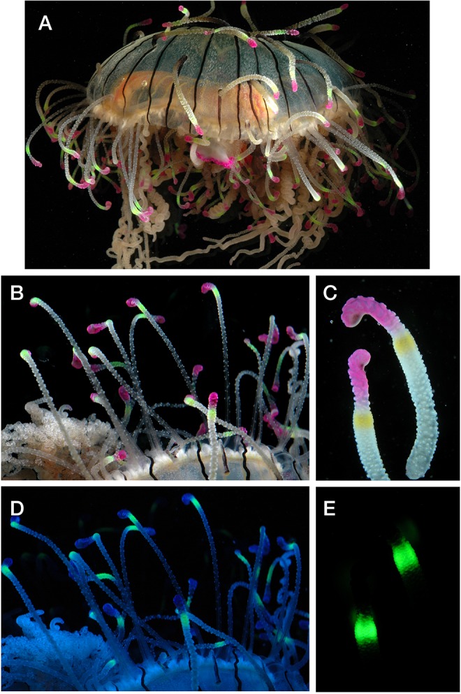
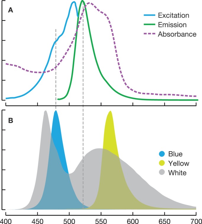
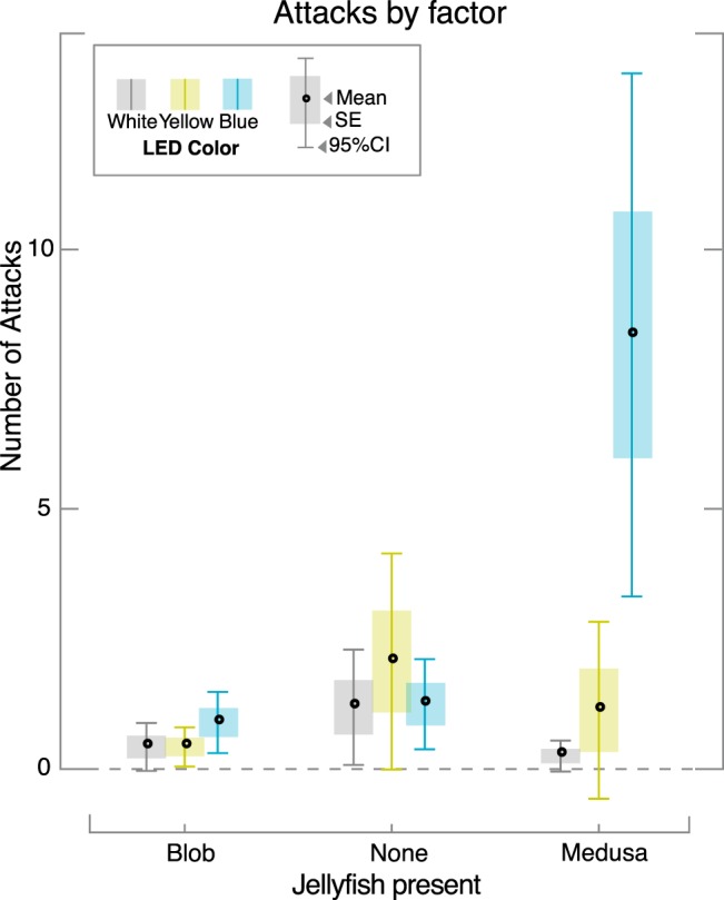
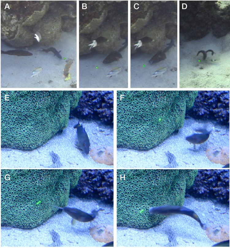
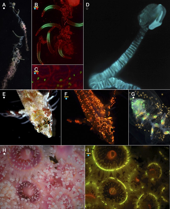
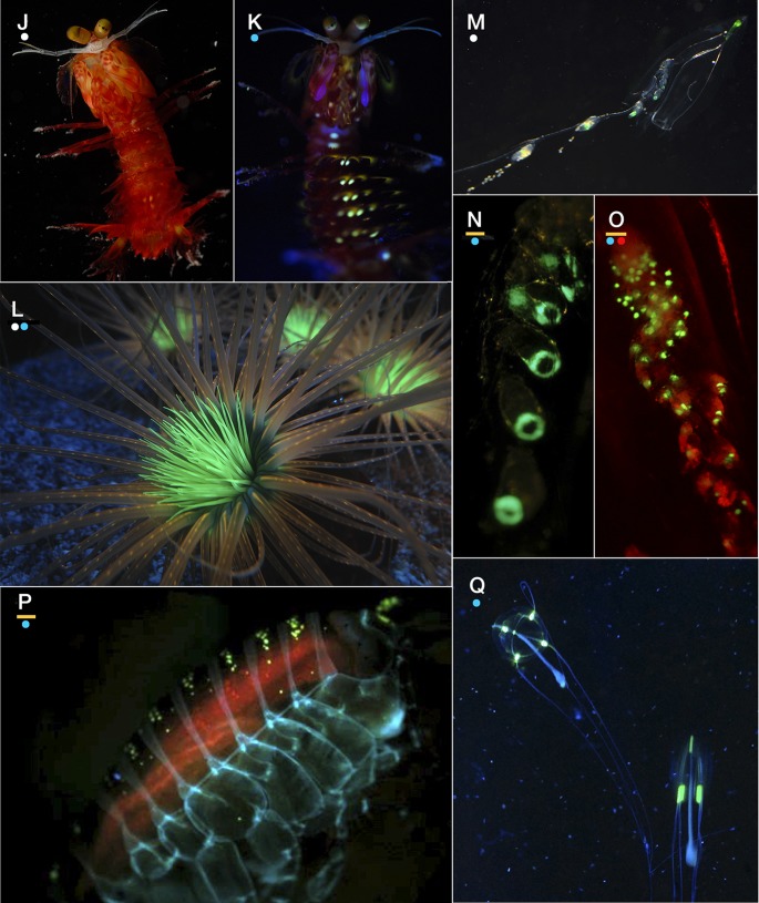
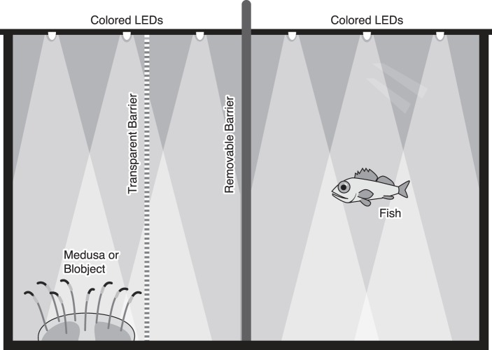
Although proteins in the green fluorescent protein family (GFPs) have been discovered in a wide array of taxa, their ecological functions in these organisms remain unclear. Many hypothesized roles are related to modifying bioluminescence spectra or modulating the light regime for algal symbionts, but these do not explain the presence of GFPs in animals that are non-luminous and non-symbiotic. Other hypothesized functions are unrelated to the visual signals themselves, including stress responses and antioxidant roles, but these cannot explain the localization of fluorescence in particular structures on the animals. Here we tested the hypothesis that fluorescence might serve to attract prey. In laboratory experiments, the predator was the hydromedusa Olindias formosus (previously known as O. formosa), which has fluorescent and pigmented patches on the tips of its tentacles. The prey, juvenile rockfishes in the genus Sebastes, were significantly more attracted (P<1×10(-5)) to the medusa's tentacles under lighting conditions where fluorescence was excited and tentacle tips were visible above the background. The fish did not respond significantly when treatments did not include fluorescent structures or took place under yellow or white lights, which did not generate fluorescence visible above the ambient light. Furthermore, underwater observations of the behavior of fishes when presented with a brightly illuminated point showed a strong attraction to this visual stimulus. In situ observations also provided evidence for fluorescent lures as supernormal stimuli in several other marine animals, including the siphonophore Rhizophysa eysenhardti. Our results support the idea that fluorescent structures can serve as prey attractants, thus providing a potential function for GFPs and other fluorescent proteins in a diverse range of organisms.

Keywords: Feeding behavior; Fluorescent protein; GFP; Olindias; Prey attraction; Supernormal stimulus.


Conflict of interest statement

Competing interests

The authors declare no competing or financial interests.

Figures

Fig. 1.
Fig. 1.
Fluorescence of Olindias. Photos of O. formosus in (A-C) white light and (D,E) under blue light, showing the fluorescence. Under white light (B) the fluorescence is excited, but is not distinct against the full-spectrum background illumination. (B,C) The tips of the tentacles have a pink chromoprotein which absorbs blue and green light, and thus appears dark in (D). Panel D is shown without a barrier filter, so the blue excitation has not been subtracted. Panel E shows the view with a long-pass filter so the blue-excitation is removed.
Fig. 2.
Fig. 2.
LED spectra subset. (A) Excitation (blue) and emission (green) of the green fluorescent protein and absorbance spectrum of the pink chromoprotein in tentacle tips of O. formosus. (B) LED emission spectra for the three treatments used in the experiment. Blue LED excites the fluorescent protein with minimal overlap with the emission spectrum (dashed grey line). Yellow LED is longer wavelength than the excitation spectrum of the fluorescence. X-axis, wavelength in nm.
Fig. 3.
Fig. 3.
Box plots of number of attacks. Number of attacks plotted by the factor (medusa present or control conditions) and for each of three lighting schemes (color of bars). Box plots show mean (dot), standard error (shaded box height) and 95% confidence interval (whisker height). Significant differences in the number of attacks (P≪1×10⁻⁵) were obtained only for the treatment that included a live medusa with blue illumination. Attack behavior with the control objects and yellow or white lighting conditions were not significantly different from each other.
Fig. 4.
Fig. 4.
Underwater housing for laser pointer used for in-situ experiments. Laser was first modified by connecting a magnetic reed switch across leads of the push-button actuator. The housing was built from plumbing hardware, using a PVC union joint which had the pipe fitting opposite the O-ring removed and replaced with a clear acrylic disk. A neodymium magnet outside the tube can be rotated to activate the reed switch inside the tube.
Fig. 5.
Fig. 5.
Frame grabs from video of the green laser deployment underwater. (A-D) Great Barrier Reef, showing wrasses pursuing the laser across the bottom and biting at it. (E-H) Aquarium footage of a goatfish responding to the appearance of the laser. Interval from E-F is 330 ms, and images G and H are each at 100 ms intervals. In image G, the barbels, laden with taste sensors, are extended to investigate the dot.
Fig. 6.
Fig. 6.
Examples of species in which fluorescence may be functioning for prey attraction. (A-C) The siphonophore Rhizophysa eysenhardti, showing white light view (A) and green fluorescence (B,C), with red illumination (not fluorescence) to show the rest of the body. (D) Bioluminescence emission of the siphonophore Rosacea plicata, with no illumination. Compare with panel G showing the distribution of fluorescence. (E,F) Light and fluorescence of the triplefin blenny Enneapterygius sp., a small tropical species with fluorescent skeletal structures. (G) White illuminated photo of Rosacea showing the fluorescence near the top of the stem and in the gastrozoids, bright enough to see without special blue excitation or filters. (H,I) White light and fluorescence of the non-symbiotic strawberry anemone Corynactis californica, showing the multi-colored fluorescence of its polyps. Scale (width of frame), A: 1.7 cm; B: 1.2 cm; C: 1.3 mm; D: 9.3 cm; E: 8.4 mm; F: 8.6 mm; G: 1.3 cm; H,I: 2.9 cm. (J,K) White light and fluorescence of the mantis shrimp Gonodactylaceus randalli. Other mantis shrimp species have strong fluorescence on their second antenna scale. (L) Cerianthid tube anemone under mixed lighting showing prominent fluorescence in central tentacles. (M-O) The siphonophore Diphyes dispar under three lighting schemes to show morphology and fluorescence associated gastrozooids (feeding polyps). Even in white light without special excitation (M) the fluorescence is visible, and it is enhanced by blue illumination (N,O). Red light in O is external illumination and not fluorescence. (P) Amphipod Cyphocaris showing several types of fluorescence: yellow from bioluminescent structure, blue from chitin, and orange likely from chlorophyll-containing gut contents. (Q) Like the hydromedusa O. formosus used in our experiments, Sarsia tubulosa has fluorescent structures that are not associated with sites of bioluminescence. Scale (width of frame), J,K: 2.9 cm; L: 9 cm; M: 2.6 cm; N: 8 mm; O: 4.7 mm; P: 11 mm; Q: 6 cm. Dots below panel letters represent color of illumination/excitation used for photos: white, blue, red, or none (bioluminescent light from organism only). Yellow bar above dots indicates when a yellow long-pass barrier filter was used.
Fig. 6.
Fig. 6.
Examples of species in which fluorescence may be functioning for prey attraction. (A-C) The siphonophore Rhizophysa eysenhardti, showing white light view (A) and green fluorescence (B,C), with red illumination (not fluorescence) to show the rest of the body. (D) Bioluminescence emission of the siphonophore Rosacea plicata, with no illumination. Compare with panel G showing the distribution of fluorescence. (E,F) Light and fluorescence of the triplefin blenny Enneapterygius sp., a small tropical species with fluorescent skeletal structures. (G) White illuminated photo of Rosacea showing the fluorescence near the top of the stem and in the gastrozoids, bright enough to see without special blue excitation or filters. (H,I) White light and fluorescence of the non-symbiotic strawberry anemone Corynactis californica, showing the multi-colored fluorescence of its polyps. Scale (width of frame), A: 1.7 cm; B: 1.2 cm; C: 1.3 mm; D: 9.3 cm; E: 8.4 mm; F: 8.6 mm; G: 1.3 cm; H,I: 2.9 cm. (J,K) White light and fluorescence of the mantis shrimp Gonodactylaceus randalli. Other mantis shrimp species have strong fluorescence on their second antenna scale. (L) Cerianthid tube anemone under mixed lighting showing prominent fluorescence in central tentacles. (M-O) The siphonophore Diphyes dispar under three lighting schemes to show morphology and fluorescence associated gastrozooids (feeding polyps). Even in white light without special excitation (M) the fluorescence is visible, and it is enhanced by blue illumination (N,O). Red light in O is external illumination and not fluorescence. (P) Amphipod Cyphocaris showing several types of fluorescence: yellow from bioluminescent structure, blue from chitin, and orange likely from chlorophyll-containing gut contents. (Q) Like the hydromedusa O. formosus used in our experiments, Sarsia tubulosa has fluorescent structures that are not associated with sites of bioluminescence. Scale (width of frame), J,K: 2.9 cm; L: 9 cm; M: 2.6 cm; N: 8 mm; O: 4.7 mm; P: 11 mm; Q: 6 cm. Dots below panel letters represent color of illumination/excitation used for photos: white, blue, red, or none (bioluminescent light from organism only). Yellow bar above dots indicates when a yellow long-pass barrier filter was used.
Fig. 7.
Fig. 7.
Experiments were conducted in a custom-built aquarium with opaque sides and transparent front. A clear barrier was fixed in place between the medusa and the fish, and an opaque barrier could be inserted between the fish and the target. The two opaque lids over the top each contained four colored LEDs, which could be changed out for the trials.

References

    1. Alieva N. O., Konzen K. A., Field S. F., Meleshkevitch E. A., Hunt M. E., Beltran-Ramirez V., Miller D. J., Wiedenmann J., Salih A. and Matz M. V. (2008). Diversity and evolution of coral fluorescent proteins. PLoS ONE 3, e2680 10.1371/journal.pone.0002680 - DOI - PMC - PubMed
    1. Arnold K. E., Owens I. P. F. and Marshall N. J. (2002). Fluorescent signaling in parrots. Science 295, 92 10.1126/science.295.5552.92 - DOI - PubMed
    1. Britt L. L., Loew E. R. and McFarland W. N. (2001). Visual pigments in the early life stages of Pacific Northwest marine fishes. J. Exp. Biol. 204, 2581-2587. - PubMed
    1. Campbell A. K. (1974). Extraction, partial purification and properties of obelin, the calcium-activated luminescent protein from the hydroid Obelia geniculata. Biochem. J. 143, 411-418. - PMC - PubMed
    1. Courtney R., Sachlikidis N., Jones R. and Seymour J. (2015). Prey capture ecology of the cubozoan Carukia barnesi. PLoS ONE 10, e0124256 10.1371/journal.pone.0124256 - DOI - PMC - PubMed