. 2015 Aug 1:12:139.
doi: 10.1186/s12974-015-0366-9.

Colony-stimulating factor 1 receptor inhibition prevents microglial plaque association and improves cognition in 3xTg-AD mice

Affiliations

Colony-stimulating factor 1 receptor inhibition prevents microglial plaque association and improves cognition in 3xTg-AD mice

Nabil N Dagher et al. J Neuroinflammation. .

Abstract

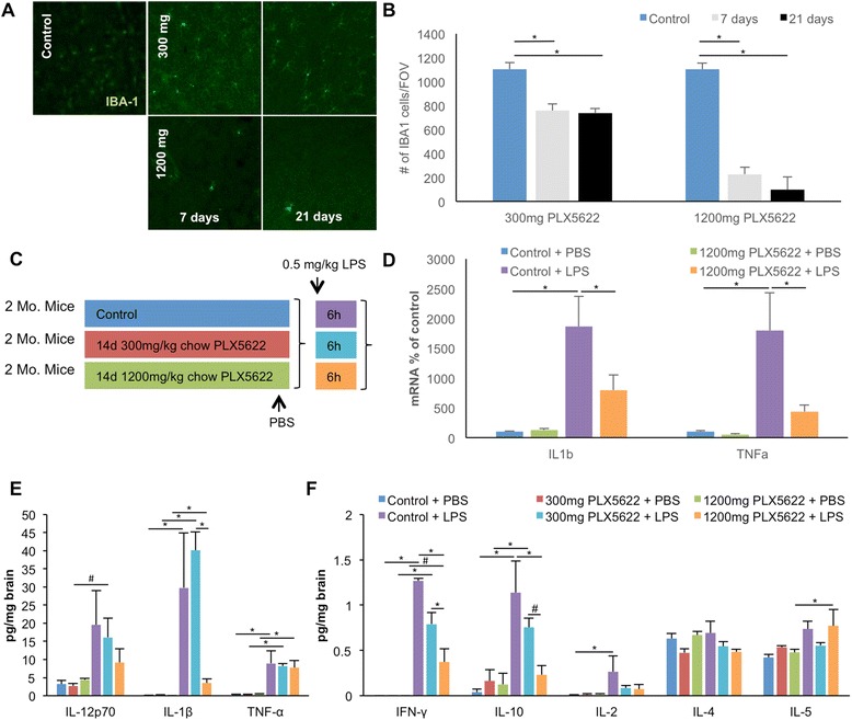
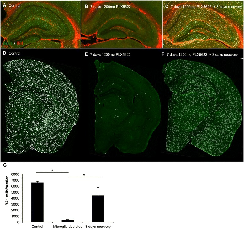
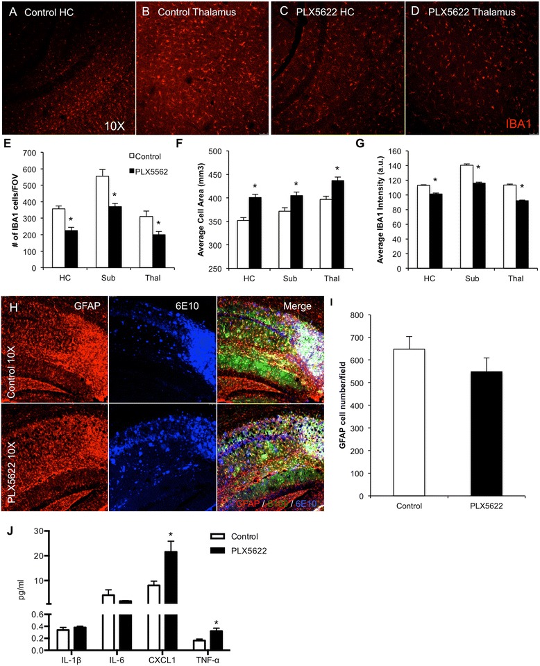
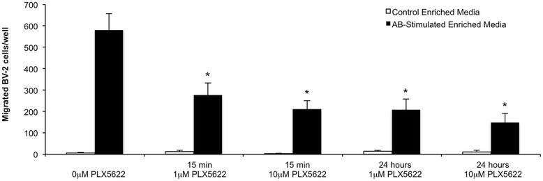
Background: Microglia are dependent upon colony-stimulating factor 1 receptor (CSF1R) signaling for their survival in the adult brain, with administration of the dual CSF1R/c-kit inhibitor PLX3397 leading to the near-complete elimination of all microglia brainwide. Here, we determined the dose-dependent effects of a specific CSF1R inhibitor (PLX5622) on microglia in both wild-type and the 3xTg-AD mouse model of Alzheimer's disease.

Methods: Wild-type mice were treated with PLX5622 for up to 21 days, and the effects on microglial numbers were assessed. 3xTg-AD mice were treated with PLX5622 for 6 or 12 weeks and effects on microglial numbers and pathology subsequently assessed.

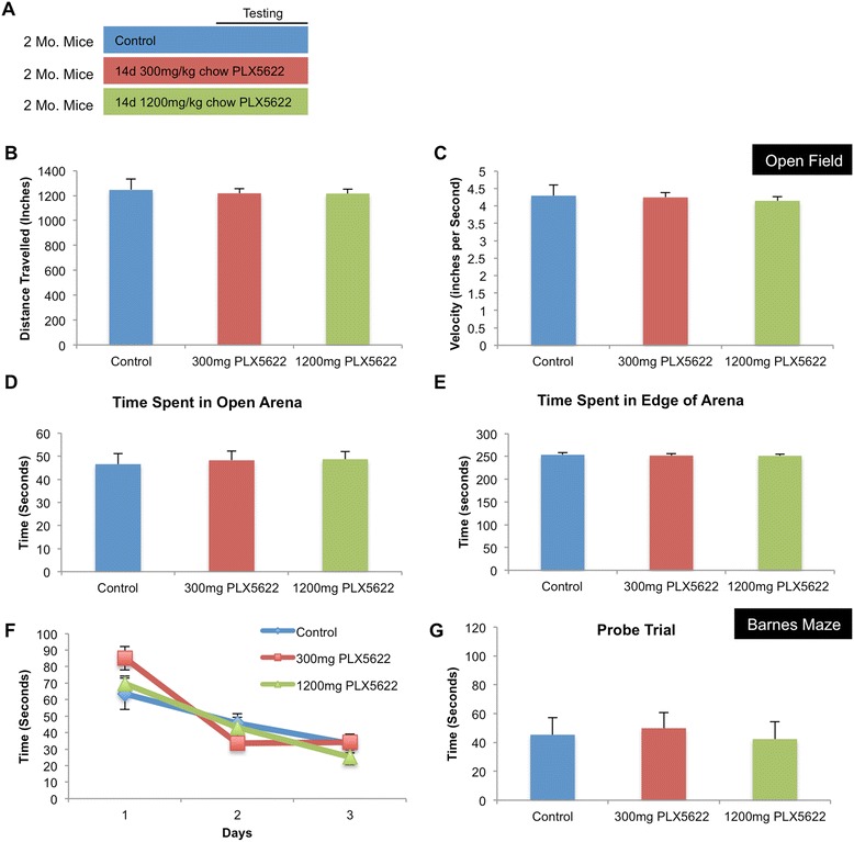
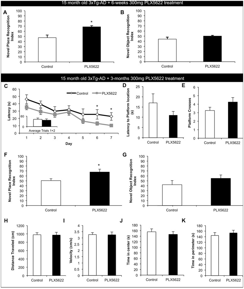
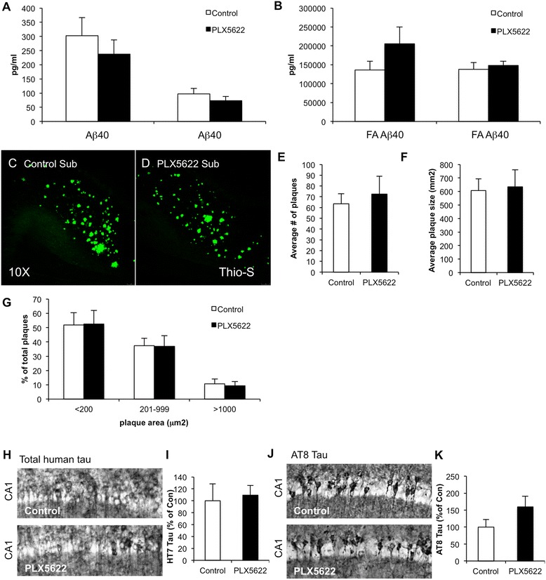
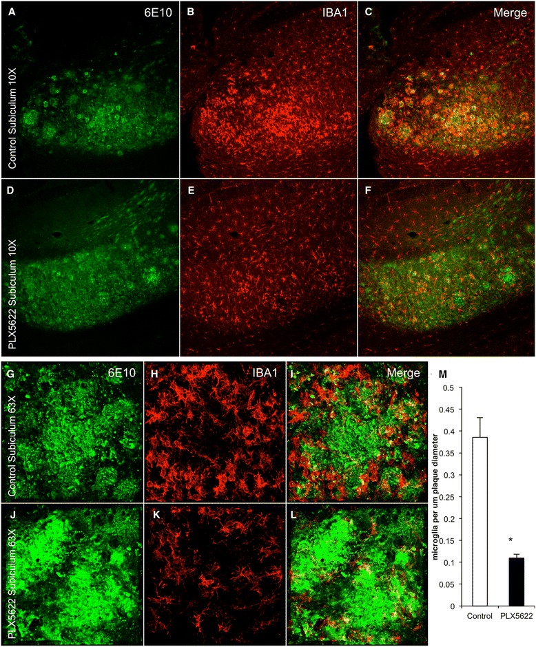
Results: High doses of CSF1R inhibitor eliminate most microglia from the brain, but a 75% lower-dose results in sustained elimination of ~30 of microglia in both wild-type and 3xTg-AD mice. No behavioral or cognitive deficits were found in mice either depleted of microglia or treated with lower CSF1R inhibitor concentrations. Aged 3xTg-AD mice treated for 6 or 12 weeks with lower levels of PLX5622 resulted in improved learning and memory. Aβ levels and plaque loads were not altered, but microglia in treated mice no longer associated with plaques, revealing a role for the CSF1R in the microglial reaction to plaques, as well as in mediating cognitive deficits.

Conclusions: We find that inhibition of CSF1R alone is sufficient to eliminate microglia and that sustained microglial elimination is concentration-dependent. Inhibition of the CSF1R at lower levels in 3xTg-AD mice prevents microglial association with plaques and improves cognition.


Figures

Fig. 1
Fig. 1
CSF1R inhibition eliminates microglia from the adult brain. Two-month-old wild-type mice were treated with vehicle, 300- or 1200 mg/kg PLX5622 for 7 or 21 days. a IBA1 immunofluorescent staining was performed on vehicle, 300 and 1200 mg/kg treated animals; representative 10× confocal images are shown for 7 and 21 days of treatment. b Quantification of the number of IBA1+ cells for all groups was performed using IMARIS software. cf 2 month-old wild-type mice were treated with 300 or 1200 mg/kg PLX5622 or vehicle for 14 days, and LPS or PBS was then administered via IP (0.5 mg/kg). mRNA for TNFα and IL-1β was measured and normalized to GAPDH in control and PLX5622-treated mice injected with PBS or LPS, showing a marked increase in both inflammatory markers in control groups injected with LPS and a dampened response to LPS in PLX5622-treated groups (d). e, f Inflammatory markers were measured via MSD® Multi-Spot Assay, revealing increases in nearly all markers with LPS injection; treatment with 1200 mg/kg PLX5622 treatment lowered the LPS-induced elevated levels of IFNγ, IL-10, and IL-1β. Treatment with 300 mg/kg PLX5622 had no significant effect on elevated levels of LPS-induced elevated levels of markers. *Indicates significance (p > 0.05), # indicates a statistical trend (p < 0.1), via two-way ANOVA with post hoc paired contrasts. Error bars indicate SEM
Fig. 2
Fig. 2
CSF1R inhibition does not alter cognition or behavior in adult mice. a Two-month-old wild-type mice were treated with either vehicle, 300 or 1200 mg/kg PLX5622 for 14 days, and behavioral analyses were conducted using an open-field test and Barnes maze. be No differences were measured across groups in the open-field test in distance traveled, velocity, time spent in open arena, or time spent in edge of arena. fg No differences were measured across groups in the Barnes maze in acquisition of time to find escape hole or in probe trial. Error bars indicate SEM
Fig. 3
Fig. 3
Microglial repopulation following microglial elimination with PLX5622 and subsequent PLX5622 withdrawal. Two-month-old wild-type mice were treated with 1200 mg/kg chow PLX5622 or vehicle for 7 days. The PLX5622 was then removed and the number of microglia was assessed 3 days later. ac IBA1/IB4 staining in control, 7 days treated with PLX5622, and 7 days treated with PLX5622 with 3 days recovery, with hippocampal region shown. df Whole brain sections were scanned and each IBA1+ cell counted. Representative brain sections are shown, with a white dot superimposed over each IBA1+ cell as a visual aid. g Quantification of df. *Indicates significance (p > 0.05) via one-way ANOVA with post hoc Newman-Keuls multiple comparison test. Error bars indicate SEM
Fig. 4
Fig. 4
Lower doses of PLX5622 improve cognition in aged 3xTg-AD mice. Fifteen-month-old 3xTg-AD mice were treated for either 6 weeks or 3 months with vehicle or 300 mg/kg PLX5622. ab Animals treated for 6 weeks were assessed using novel place and novel object recognition tasks. Treated mice showed a significant improvement in place recognition as compared to untreated 3xTg-AD mice, but no differences were measured between groups in novel object recognition. ck Animals treated for 3 months were assessed using Morris water maze, novel place recognition, and novel object recognition. ce Morris water maze. Treated mice had significantly faster escape latencies on days 6 and 7 of Morris water maze training and trended towards a faster latency to reach platform and increased platform crosses during the probe trial. f, g Treated mice showed significantly improved place recognition as compared to control mice, but no differences were shown between groups in novel object recognition. hk No differences in open field were detected in either distance traveled (h), average velocity (i), the time spent in the center of the arena (j), or in the time spent in the perimeter of the arena (k). *Indicates significance (p < 0.05) by unpaired Student’s t test. Error bars indicate SEM
Fig. 5
Fig. 5
Lower-dose CSF1R inhibition partially reduces microglia numbers. The brains of 3 months 3xTg-AD-treated mice were examined for effects of PLX5622 on pathology. ad IBA1 immunofluorescent staining was performed and representative 10× images are shown of control and treated hippocampus and thalamus. e IBA1+ cell counts revealed a reduction by 30 % in the treated groups. f, g IBA1+ cells in treated brains are larger but have reduced staining intensity as compared to 3xTg-AD untreated mice. h Immunofluorescent staining for the astrocytic markers GFAP (red), S100 (green), and plaques with 6E10 (blue), with the hippocampal region shown. i Quantification of the number of GFAP+ cells in the hippocampal sub-field. j Inflammatory profiling of whole brain homogenates shows significant increases in CXCL1 and TNFα but not Il-1β or Il-6. *Indicates significance (p < 0.05) by unpaired Students t test. Error bars indicate SEM
Fig. 6
Fig. 6
Lower-dose CSF1R inhibition does not alter Aβ or Tau levels. a, b Sandwich ELISA in soluble and insoluble fraction revealed no significant differences in Aβ40 or Aβ42 between groups. c, d Thioflavin-S staining was performed, revealing plaques in both control and treated groups. Of the subiculum, 10× images are shown. eg Analysis of plaques revealed no significant difference between treated and untreated brains in average number of plaques, average plaque size, or distribution of large, medium, and small plaques. hk Immunostaining for total human tau with HT7 (h, i) and AT8 tau (j, k) reveals no significant differences in tau levels with PLX5622 treatment. Error bars indicate SEM
Fig. 7
Fig. 7
Chronic lower-dose CSF1R inhibition prevents microglia associating with plaques. Immunofluorescent staining was performed on 3 months treated and control 3xTg-AD mice for 6E10, which recognizes Aβ plaques and IBA1. af Representative 10× images are shown of control and treated mice. gl Representative 63× images are shown of control and treated mice, centered on an area dense with plaques. m Quantification of the number of microglia associated with a plaque and normalized to plaque diameter revealed a 70 % decrease in treated animals as compared to untreated animals. *Indicates significance (p < 0.05) by unpaired Students t test. Error bars indicate SEM
Fig. 8
Fig. 8
PLX5622 inhibits chemotaxis of BV-2 cells in response to Aβ-oligomer-stimulated enriched media. Chemotaxis was measured by counting migrated BV2 cells in response to Aβ-stimulated enriched media or control enriched media. BV2 cells were treated with 0-, 1-, or 10 μM PLX5622 either 15 min or 24 h before the assay was conducted. All treated cells exhibited significantly reduced cell migration in response to the Aβ-stimulated enriched media

References

    1. Ramlackhansingh AF, Brooks DJ, Greenwood RJ, Bose SK, Turkheimer FE, Kinnunen KM, et al. Inflammation after trauma: microglial activation and traumatic brain injury. Ann Neurol. 2011;70(3):374–83. doi: 10.1002/ana.22455. - DOI - PubMed
    1. Wang Q, Tang XN, Yenari MA. The inflammatory response in stroke. J Neuroimmunol. 2007;184(1–2):53–68. doi: 10.1016/j.jneuroim.2006.11.014. - DOI - PMC - PubMed
    1. Lenzlinger PM, Morganti-Kossmann MC, Laurer HL, McIntosh TK. The duality of the inflammatory response to traumatic brain injury. Mol Neurobiol. 2001;24(1–3):169–81. - PubMed
    1. Wyss-Coray T, Lin C, Yan F, Yu GQ, Rohde M, McConlogue L, et al. TGF-beta1 promotes microglial amyloid-beta clearance and reduces plaque burden in transgenic mice. Nat Med. 2001;7(5):612–8. doi: 10.1038/87945. - DOI - PubMed
    1. Rice RA, Yamate-Morgan H, Lee RJ, Arora RPS, Hernandez MX, Tenner AJ, et al. Elimination of microglia improves functional outcomes following extensive neuronal loss in the hippocampus. J Neurosci. 2015;35(27):9977–89. - PMC - PubMed

Publication types

MeSH terms

Substances