. 2015 Sep 10;126(11):1357-66.
doi: 10.1182/blood-2015-01-624809. Epub 2015 Jul 31.

Transcriptome analysis highlights the conserved difference between embryonic and postnatal-derived alveolar macrophages

Affiliations

Transcriptome analysis highlights the conserved difference between embryonic and postnatal-derived alveolar macrophages

Sophie L Gibbings et al. Blood. .

Abstract

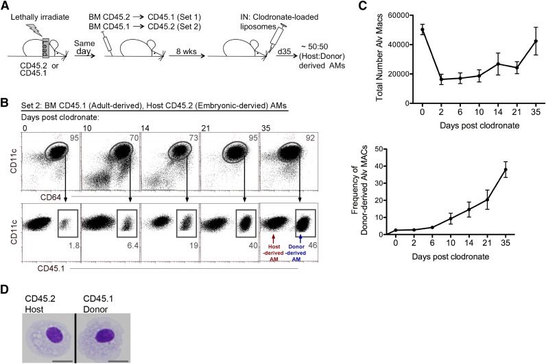
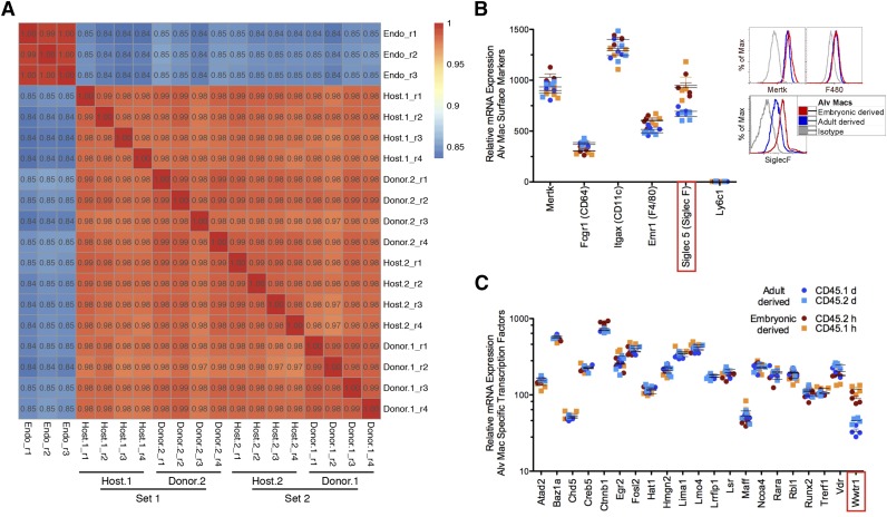
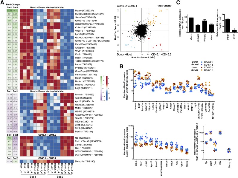
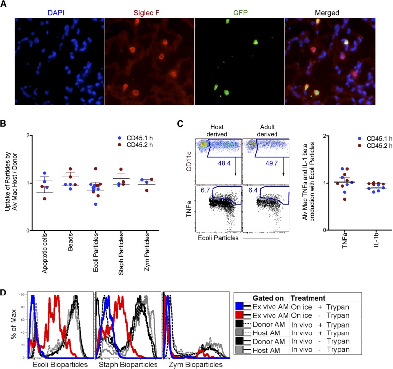
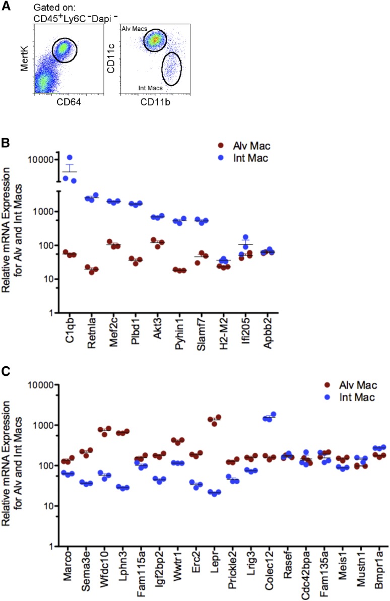
Alveolar macrophages (AMs) reside on the luminal surfaces of the airways and alveoli where they maintain host defense and promote alveolar homeostasis by ingesting inhaled particulates and regulating inflammatory responses. Recent studies have demonstrated that AMs populate the lungs during embryogenesis and self-renew throughout life with minimal replacement by circulating monocytes, except under extreme conditions of depletion or radiation injury. Here we demonstrate that on a global scale, environment appears to dictate AM development and function. Indeed, transcriptome analysis of embryonic host-derived and postnatal donor-derived AMs coexisting within the same mouse demonstrated >98% correlation and overall functional analyses were similar. However, we also identified several genes whose expression was dictated by origin rather than environment. The most differentially expressed gene not altered by environment was Marco, a gene recently demonstrated to have enhancer activity in embryonic-derived but not postnatal-derived tissue macrophages. Overall, we show that under homeostatic conditions, the environment largely dictates the programming and function of AMs, whereas the expression of a small number of genes remains linked to the origin of the cell.


Figures

Figure 1
Figure 1
Model for the coexistence of host- and donor-derived AMs. (A) Strategy: First, lungs of mice were protected with lead prior to irradiation. Postirradiation, congenic BM cells were transferred intravenously. Set 1: irradiated CD45.1 mice received CD45.2 BM. Set 2: irradiated CD45.2 mice received CD45.1 BM. Eight weeks later, mice were treated with CLL via intranasal (IN) delivery to deplete AMs. By day 35, post-CLL treatment, mice contained ∼50% host- or donor-derived AMs. (B) Time course analysis of the reconstitution of host- and donor-derived AMs post-CLL treatment. Frequency of contribution of CD45.1+ donor-derived CD11c+CD64+ AMs was analyzed (illustrated by set 2). Data represents 3 independent experiments. (C) Time course analysis of the total numbers of AM post-CLL treatment. Data represents 3 independent experiments. (D) Morphologic analysis of BM reconstituted mice at day 35 post-CLL. Cytospin of sorted host- and donor-derived AM. MACs, macrophages.
Figure 2
Figure 2
Transcriptional profiles of host- and donor-derived AMs. For microarray analysis, AMs of host and donor origin were fluorescence-activated cell sorted from whole lung digests from lead shielded BM reconstituted mice 8-weeks post-CLL delivery. (A) Pearson correlation plots comparing host- and donor-derived AMs from either CD45.1 (host.1 and donor.1) or CD45.2 (host.2 and donor.2). Data demonstrates experimental sets containing 4 replicates (r) of each sample type. An endothelial cell line (Bend3) was used as a control for correlation comparison (Endo). (B) Messenger RNA (mRNA) microarray expression for surface molecules on host-derived AMs (red, CD45.2 host; orange, CD45.1 host) and donor-derived AMs (blue, CD45.1 donor; light blue, CD45.2 donor) (left). Protein expression by flow cytometry for Siglec F, MertK, and F4/80 on host- and donor-derived AMs (right). Protein data represents 8 independent experiments. (C) The mRNA microarray expression levels of lung specific transcription factors. Statistically significant differences (Student t test; P < .05) between host- and donor-derived AMs are highlighted in red box. Alv Macs, alveolar macrophages.
Figure 3
Figure 3
Differences in gene expression between host- and donor-derived AMs. (A) Heatmap illustrating expression level profiles for gene with a significant (limma, false discovery rate = 0.05) difference for host vs donor-derived AM or CD45.1 vs CD45.2-derived AM) and at least a twofold change difference between host- and donor-derived AMs in both set 1 (host.1 vs donor.2) and set 2 (host.2 vs donor.1) (left). Probe identifiers are shown in parentheses after gene symbols annotating each row. Row-scaled expression levels shown using red-blue scale, fold-change shown using green-purple scale. Right, four-way scatter plot comparing fold change in set 1 and fold change in set 2. Genes highlighted in orange, blue, red, or green represent the 4 possible extreme groups host > donor, host < donor, CD45.1 > CD45.2, and CD45.1 < CD45.2, respectively, whose data were shown in the heatmap (left). Gray dashed lines depict a twofold change difference. (B) Scatter plot of relative expression of messenger RNA (mRNA) for genes represented on heat map. (C) Confirmation of Marco, Sema3e, Wfdc10, Wwtr1, Pldb1, and Ifi205 microarray gene expression by quantitative PCR. Data are representative of 3 independent experiments.
Figure 4
Figure 4
Functional analysis of host- and donor-derived AMs. (A) Microscopy staining around the alveolus for Siglec F+ host-derived (GFP) and donor-derived (GFP+) AMs was performed on lead shielded BM reconstituted mice 8-weeks post-CLL delivery. (B) Ratio analysis of host- over donor-derived AMs uptake for apoptotic cell, carboxylated beads, and bioparticles, 2 hours postintranasal delivery. (C) Flow cytometry gating strategy for 4B-4C (left) and ratio analysis of host- over donor-derived AMs for TNF-α and IL-1β production from AM with ingested Escherichia coli (right). Each dot represents one mouse. Blue dots represents analysis from set 1 and red dots represent analysis from set 2 (set 1 and set 2 as described in Figure 1A). (D) Two hours post-IN delivery of bioparticles, AMs were lavaged and quenched to exclude surface bound bioparticles. Black and gray lines are donor- and host-derived AM, dashed is quenched; solid line is unquenched. Control quenching was assessed with ex vivo AM given bioparticles on ice. Blue line is quenched; red line is unquenched. DAPI, 4,6 diamidino-2-phenylindole; GFP, green fluorescent protein.
Figure 5
Figure 5
Selective gene analysis of alveolar and interstitial macrophages from naïve mice. (A) Identification CD45+ (Lin: CD3 B220 NK1.1 Ly6C Ly6G) CD64+MertK+, CD11c+CD11b alveolar and CD11cCD11b+ interstitial macrophages. (B) Scatter plot of relative expression of messenger RNA (mRNA) for genes selectively expressed in favor of donor-derived AMs from alveolar (red) and interstitial (blue) macrophages isolated from naïve mice. Data shows triplicate sorts. (C) Scatter plot of relative expression of mRNA for genes selectively expressed in favor of host-derived AMs from alveolar (red) and interstitial (blue) macrophages isolated from naïve mice. Data illustrates triplicate sorts. Int Mac, interstitial macrophages.

Similar articles

Cited by

References

    1. MacLean JA, Xia W, Pinto CE, Zhao L, Liu HW, Kradin RL. Sequestration of inhaled particulate antigens by lung phagocytes. A mechanism for the effective inhibition of pulmonary cell-mediated immunity. Am J Pathol. 1996;148(2):657–666. - PMC - PubMed
    1. Thepen T, Van Rooijen N, Kraal G. Alveolar macrophage elimination in vivo is associated with an increase in pulmonary immune response in mice. J Exp Med. 1989;170(2):499–509. - PMC - PubMed
    1. Thepen T, Kraal G, Holt PG. The role of alveolar macrophages in regulation of lung inflammation. Ann N Y Acad Sci. 1994;725:200–206. - PubMed
    1. Guilliams M, Lambrecht BN, Hammad H. Division of labor between lung dendritic cells and macrophages in the defense against pulmonary infections. Mucosal Immunol. 2013;6(3):464–473. - PubMed
    1. Gautier EL, Shay T, Miller J, et al. Immunological Genome Consortium. Gene-expression profiles and transcriptional regulatory pathways that underlie the identity and diversity of mouse tissue macrophages. Nat Immunol. 2012;13(11):1118–1128. - PMC - PubMed

Publication types

MeSH terms