. 2015 Oct;1852(10 Pt A):2225-2234.
doi: 10.1016/j.bbadis.2015.07.019. Epub 2015 Jul 29.

NR2B-dependent cyclophilin D translocation suppresses the recovery of synaptic transmission after oxygen-glucose deprivation

Affiliations

NR2B-dependent cyclophilin D translocation suppresses the recovery of synaptic transmission after oxygen-glucose deprivation

Zhihua Zhang et al. Biochim Biophys Acta. 2015 Oct.

Abstract

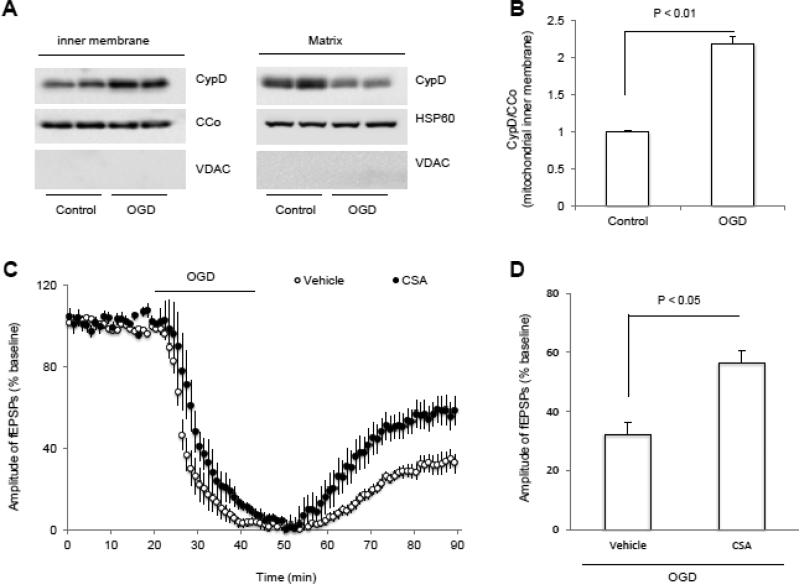
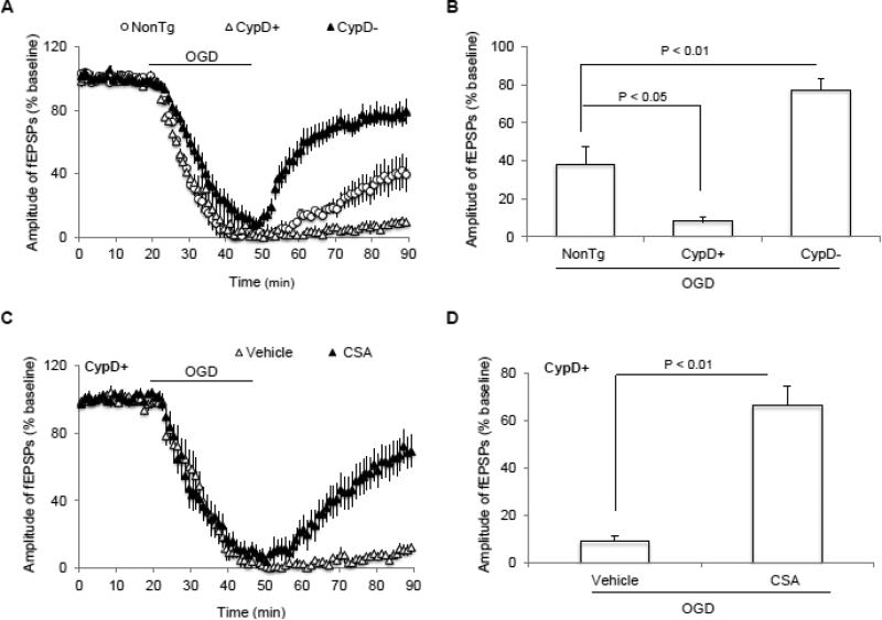
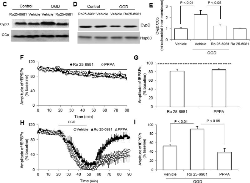
N-methyl d-aspartate receptor (NMDA) subunit 2B (NR2B)-containing NMDA receptors and mitochondrial protein cyclophilin D (CypD) are well characterized in mediating neuronal death after ischemia, respectively. However, whether and how NR2B and CypD work together in mediating synaptic injury after ischemia remains elusive. Using an ex vivo ischemia model of oxygen-glucose deprivation (OGD) in hippocampal slices, we identified a NR2B-dependent mechanism for CypD translocation onto the mitochondrial inner membrane. CypD depletion (CypD null mice) prevented OGD-induced impairment in synaptic transmission recovery. Overexpression of neuronal CypD mice (CypD+) exacerbated OGD-induced loss of synaptic transmission. Inhibition of CypD-dependent mitochondrial permeability transition pore (mPTP) opening by cyclosporine A (CSA) attenuated ischemia-induced synaptic perturbation in CypD+ and non-transgenic (non-Tg) mice. The treatment of antioxidant EUK134 to suppress mitochondrial oxidative stress rescued CypD-mediated synaptic dysfunction following OGD in CypD+ slices. Furthermore, OGD provoked the interaction of CypD with P53, which was enhanced in slices overexpressing CypD but was diminished in CypD-null slices. Inhibition of p53 using a specific inhibitor of p53 (pifithrin-μ) attenuated the CypD/p53 interaction following OGD, along with a restored synaptic transmission in both non-Tg and CypD+ hippocampal slices. Our results indicate that OGD-induced CypD translocation potentiates CypD/P53 interaction in a NR2B dependent manner, promoting oxidative stress and loss of synaptic transmission. We also evaluate a new ex vivo chronic OGD-induced ischemia model for studying the effect of oxidative stress on synaptic damage.

Keywords: CypD; Mitochondria; NR2B; OGD; Synaptic transmission; p53.


Figures

Figure 1
Figure 1. Identification and characterization of transgenic (Tg) CypD+ mice
A) Cyclophilin D (CypD) transgenic mice (+) and nontransgenic (nonTg, −) control mice were identified by PCR results. B-C) Immunoblotting of brain homogenates from Tg CypD (lane 2, +) mice and nonTg littermate controls (lane 1, −) for CypD, using anti-human CypD antibody. C) Quantification of CypD immunoreactive bands normalized to β-actin. Data are presented as fold increase relative to nonTg mice. N = 5-6 mice/group. D-F). The double immunofluorescent staining of brain sections for CypD (red) and MAP2 (green) in hippocampus (D) and cortex (E) from the indicated Tg mice. Nuclei were stained by DRAQ5 as shown in blue. F) Quantification of CypD staining intensity in hippocampus and cortex regions of the indicated Tg mice. G) Representative immunostaining images for CypD (green) and SODII (red, mitochondrial marker) and nuclei (blue) in hippocampal and cortical neurons. Scale bar = 25 μm.
Figure 2
Figure 2. OGD triggers CypD translocation and inhibition of mPTP by cyclosporin A (CSA) promotes synaptic transmission recovery after OGD
A) Representative immunoblotting bands show CypD levels in mitochondrial inner membrane in the indicated groups of slices. VDAC, CCo and HSP60 were used as out membrane of mitochondria, inner membrane of mitochondria and mitochondrion matrix marker, respectively. B) Quantification of CypD immunoactive bands relative to CCo in the indicated groups. N = 4 mice per group. C) Changes of the amplitude of field-excitatory post-synaptic potentials (fEPSPs) in indicated groups. CSA (1 μM) treatment started 5 min before OGD (bar) and presented during entire OGD period. D) Synaptic transmission recovery of fEPSPs calculated as the averaged relative amplitude of fEPSPs compared to baseline values after re-introduction of oxygenated normal ACSF (from 35 to 40 min after the end of OGD). N =9 slices from 4-5 male mice (3-4 month-old age) per group.
Figure 3
Figure 3. Effect of CypD on synaptic dysfunction after ischemia
A) Cyclophilin D (CypD) overexpression suppresses and CypD deficiency restores synaptic transmission after oxygen and glucose deprivation (OGD), respectively. B) Summary of the field-excitatory post-synaptic potentials (fEPSPs) recovery during the last 5 min of OGD in indicated groups. C) Inhibition of CypD− mPTP by cyclosporin A (CSA) during OGD significantly preserved synaptic transmission in CypD overexpressed animals. D) Synaptic transmission recovery of fEPSPs calculated as the averaged relative amplitude of fEPSPs compared to baseline values after re-introduction of oxygenated normal artificial cerebrospinal fluid (ACSF, see methods section) (from 35 to 40 min after the end of OGD) in indicated groups. N = 8-14 slices from 4-5 male mice per group.
Figure 4
Figure 4. N-methyl D-aspartate receptor subunit 2B (NR2B) activation is involved in OGD-induced cyclophilin D (CypD) translocation and synaptic injury
A-B) Representative immunoblotting bands (A) and Quantification of (B) show the phosphorylation level of NR2B at ser1303 in indicated groups. C-D) Representative immunoblotting bands show CypD levels in mitochondrial inner membrane fraction and matrix (D) in indicated groups. E) Quantification of CypD immunoreactive bands normalized to CCo in indicated groups shown in panel C. N =4 mice per group. F) Inhibition of either NR2A (PPPA, 0.5 μM) or NR2B (Ro 25-6981, 1 μM) by its inhibitor perfusion (bar) suppressed synaptic transmission under normal condition. G) Average of the last 5 min of reperfusion fEPSPs amplitude in the indicated groups. H) Inhibition of NR2B but not NR2A significantly ameliorated synaptic injury after OGD. I) Synaptic transmission recovery of field-excitatory post-synaptic potentials (fEPSPs) calculated as the averaged relative amplitude of fEPSPs compared to baseline values after re-introduction of oxygenated normal artificial cerebrospinal fluid (ACSF, see methods section) (from 35 to 40 min after the end of OGD). N = 6-10 slices from 4-5 male mice (3-4 month-old age) per group.
Figure 5
Figure 5. The effect of p53 and NR2B activation on OGD-induced CypD/p53 complex formation in nonTg mice
A) Immunoprecipitation of hippocampal homogenates with p53 antibody followed by immunoblotting with CypD antibody revealed CypD immunoreactive bands at 53kD in OGD-exposed non-transgenic (nonTg) hippocampal tissue. CypD/p53 complex was elevated in CypD-overexpressed mice and absent in CypD-deficient animals. β-actin bands show the equal amounts of protein used for Co-immunoprecipitation experiments. B). Pifithrin-μ (PFT, 5μM) treatment significantly suppressed OGD-induced CypD/p53 interaction in mitochondrial fractions. C) Inactivation of NR2B subunit instead of NR2A prevented CypD/p53 interaction from the isolated brain mitochondria after OGD. Experiments repeated at least 3 times; 5-6 mice per group. PFT: pifithrin-μ; Ro25: Ro25-6981
Figure 6
Figure 6. Suppression of CypD/p53 complex formation via blockade of p53 or antioxidant EUK134 perfusion maintained synaptic function after oxygen and glucose deprivation (OGD)
A) p53 inhibitor, pifithrin-μ (PFT, 5 μM), perfusion (dash line) of non-transgenic (nonTg) slices did not change field-excitatory post-synaptic potentials (fEPSPs) under baseline conditions. However, it significantly promoted synaptic transmission recovery after OGD (solid bar). B) Synaptic transmission recovery of fEPSPs calculated as the averaged relative amplitude of fEPSPs compared to baseline values after re-introduction of oxygenated normal artificial cerebrospinal fluid (from 35 to 40 min after the end of OGD). C-D) CypD-overexpressed slices pretreated with p53 inhibitor preserved synaptic transmission after OGD. E-F) CypD/p53 complex formation increases due to oxidative stress. The antioxidant EUK134 pretreatment (0.5 μM) abolishes the deleterious effect of OGD on synaptic transmission in CypD+ slices. N = 6-10 slices from 4-5 male mice (3-4 month-old age) per group.

Similar articles

Cited by

References

    1. Albensi BC, Igoechi C, Janigro D, Ilkanich E. Why do many NMDA antagonists fail, while others are safe and effective at blocking excitotoxicity associated with dementia and acute injury? Am J Alzheimers Dis Other Demen. 2004;19:269–274. - PMC - PubMed
    1. Arundine M, Tymianski M. Molecular mechanisms of glutamate-dependent neurodegeneration in ischemia and traumatic brain injury. Cell Mol Life Sci. 2004;61:657–668. - PMC - PubMed
    1. Baines CP, Kaiser RA, Purcell NH, Blair NS, Osinska H, Hambleton MA, Brunskill EW, Sayen MR, Gottlieb RA, Dorn GW, Robbins J, Molkentin JD. Loss of cyclophilin D reveals a critical role for mitochondrial permeability transition in cell death. Nature. 2005;434:658–662. - PubMed
    1. Cao J, Viholainen JI, Dart C, Warwick HK, Leyland ML, Courtney MJ. The PSD95-nNOS interface: a target for inhibition of excitotoxic p38 stress-activated protein kinase activation and cell death. The Journal of cell biology. 2005;168:117–126. - PMC - PubMed
    1. Chan PH. Mitochondria and neuronal death/survival signaling pathways in cerebral ischemia. Neurochemical research. 2004;29:1943–1949. - PubMed

LinkOut - more resources