. 2015 Aug 1;19(2):pyv091.
doi: 10.1093/ijnp/pyv091.

Impaired 2-AG Signaling in Hippocampal Glutamatergic Neurons: Aggravation of Anxiety-Like Behavior and Unaltered Seizure Susceptibility

Affiliations

Impaired 2-AG Signaling in Hippocampal Glutamatergic Neurons: Aggravation of Anxiety-Like Behavior and Unaltered Seizure Susceptibility

Stephan Guggenhuber et al. Int J Neuropsychopharmacol. .

Erratum in

  • Erratum.
    [No authors listed] [No authors listed] Int J Neuropsychopharmacol. 2016 Apr 27;19(10):pyw031. doi: 10.1093/ijnp/pyw031. Int J Neuropsychopharmacol. 2016. PMID: 27207904 Free PMC article. No abstract available.

Abstract

Background: Postsynaptically generated 2-arachidonoylglycerol activates the presynaptic cannabinoid type-1 receptor, which is involved in synaptic plasticity at both glutamatergic and GABAergic synapses. However, the differential function of 2-arachidonoylglycerol signaling at glutamatergic vs GABAergic synapses in the context of animal behavior has not been investigated yet.

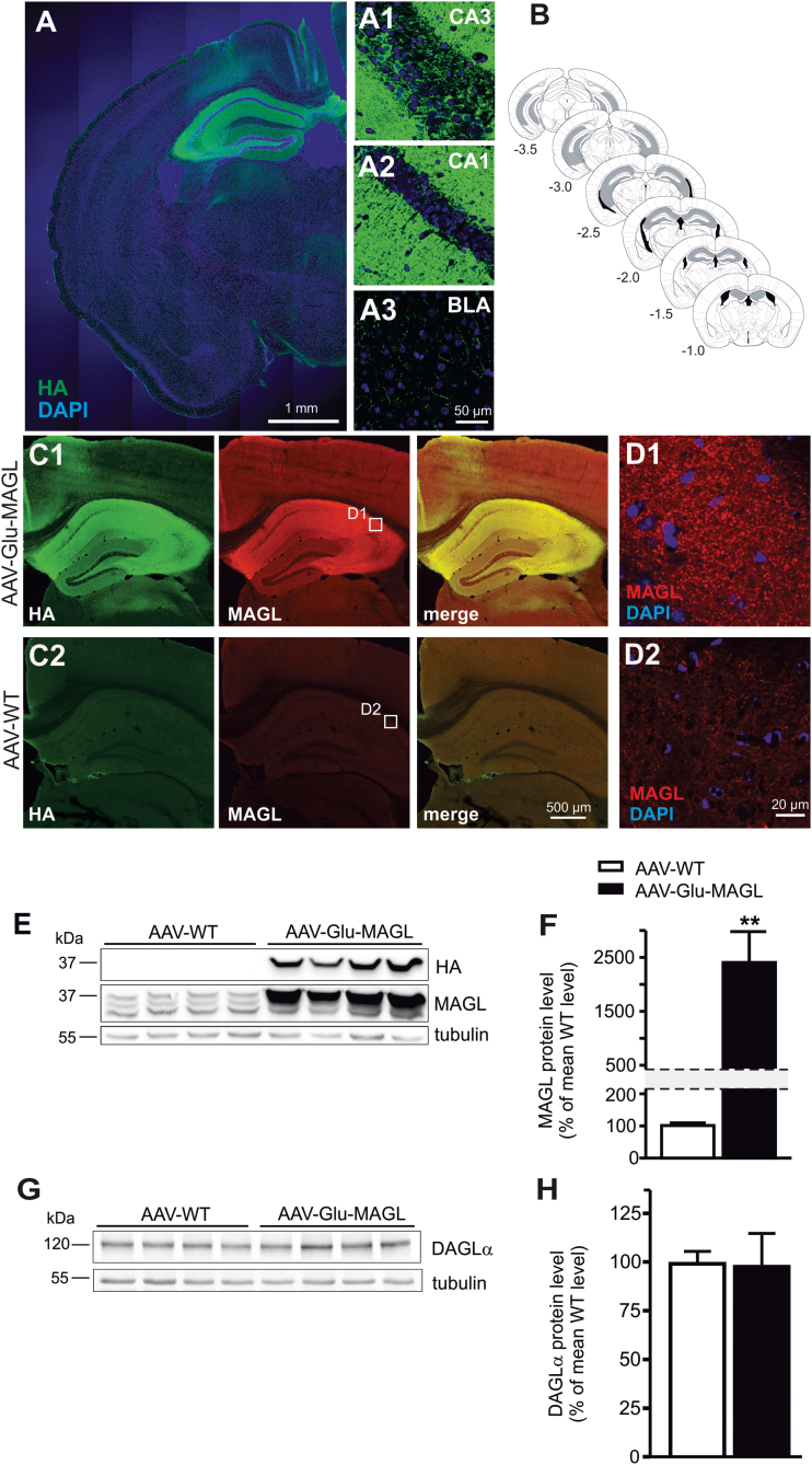
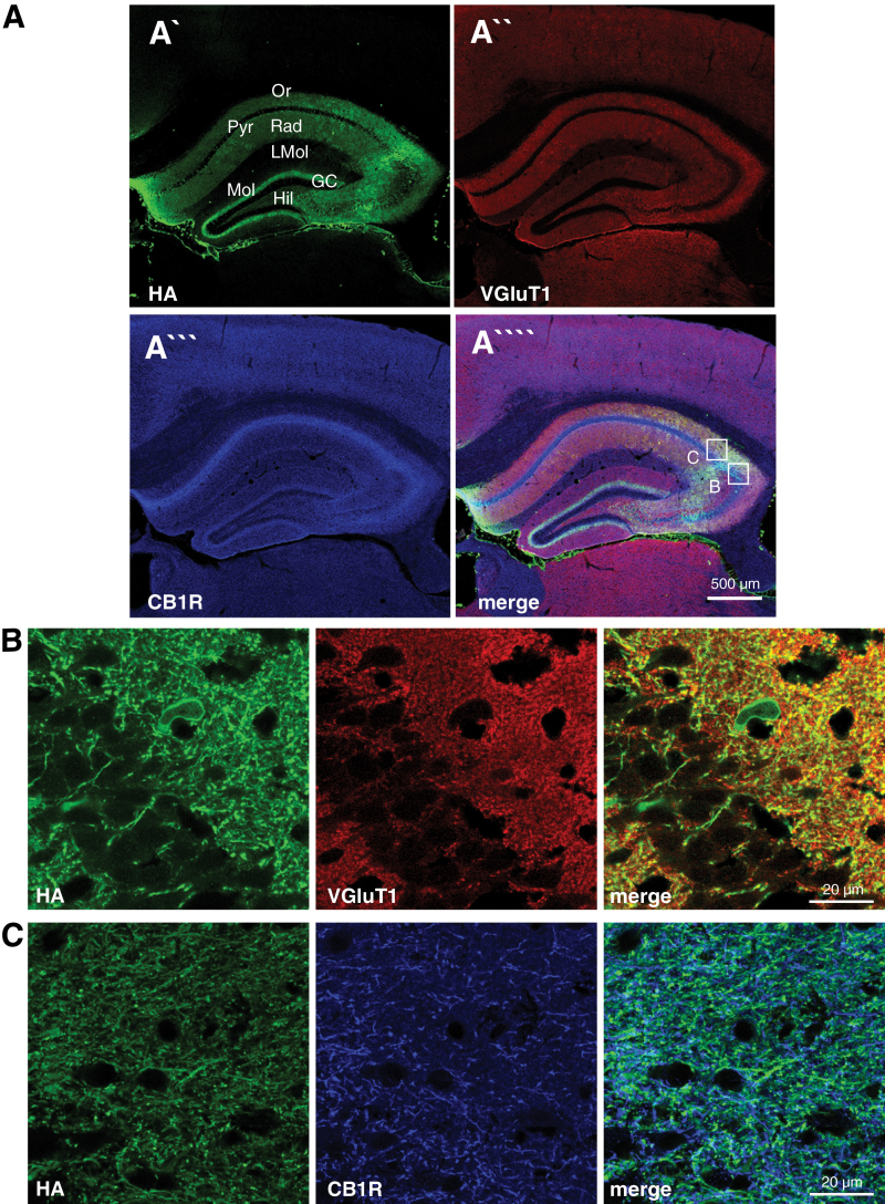
Methods: Here, we analyzed the role of 2-arachidonoylglycerol signaling selectively in hippocampal glutamatergic neurons. Monoacylglycerol lipase, the primary degrading enzyme of 2-arachidonoylglycerol, is expressed at presynaptic sites of excitatory and inhibitory neurons. By adeno-associated virus-mediated overexpression of monoacylglycerol lipase in glutamatergic neurons of the mouse hippocampus, we selectively interfered with 2-arachidonoylglycerol signaling at glutamatergic synapses of these neurons.

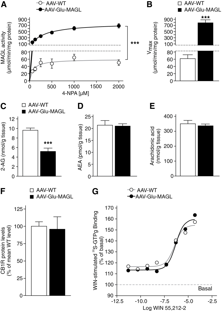
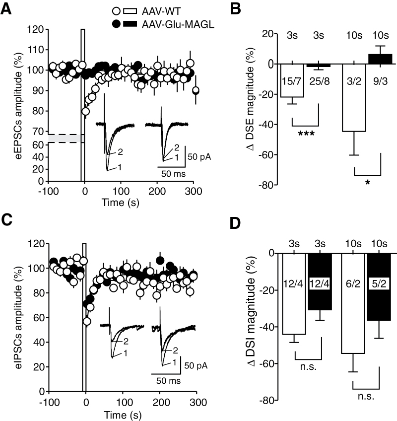
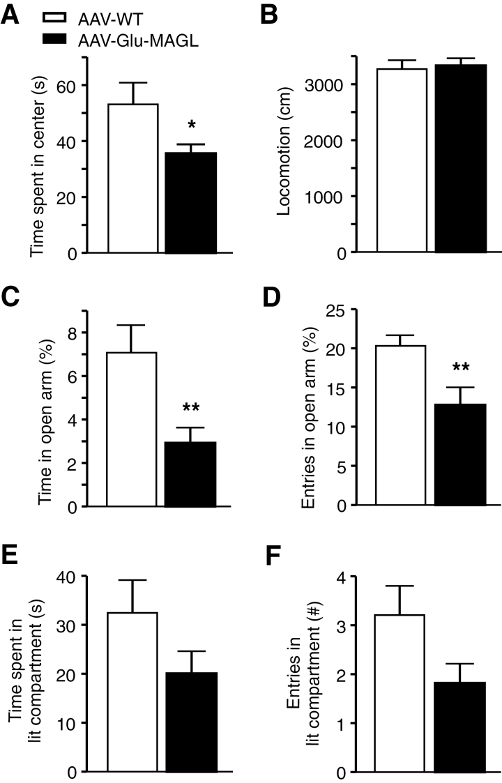
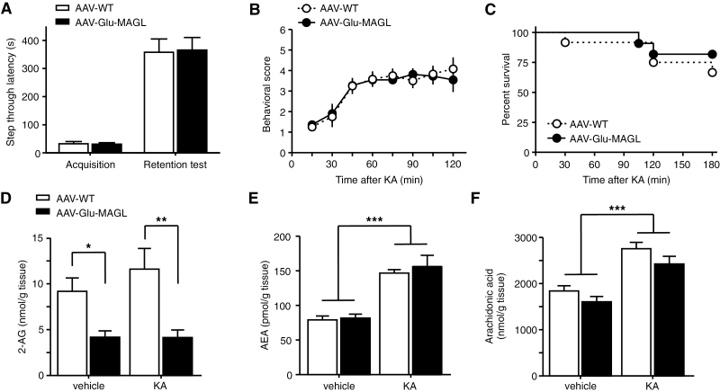
Results: Genetic modification of monoacylglycerol lipase resulted in a 50% decrease in 2-arachidonoylglycerol tissue levels without affecting the content of the second major endocannabinoid anandamide. A typical electrophysiological read-out for 2-arachidonoylglycerol signaling is the depolarization-induced suppression of excitation and of inhibition. Elevated monoacylglycerol lipase levels at glutamatergic terminals selectively impaired depolarization-induced suppression of excitation, while depolarization-induced suppression of inhibition was not significantly changed. At the behavioral level, mice with impaired hippocampal glutamatergic 2-arachidonoylglycerol signaling exhibited increased anxiety-like behavior but showed no alterations in aversive memory formation and seizure susceptibility.

Conclusion: Our data indicate that 2-arachidonoylglycerol signaling selectively in hippocampal glutamatergic neurons is essential for the animal's adaptation to aversive situations.

Keywords: Monoacylglycerol lipase; anxiety; endocannabinoids; epilepsy; hippocampus.


Figures

Figure 1.
Figure 1.
Monoacylgycerol lipase (MAGL) overexpression in hippocampal pyramidal neurons. (A) Overview representation of a section of one brain hemisphere, showing hemagglutinin (HA) immunostaining and indicating strong MAGL transgene expression in hippocampal pyramidal neurons of adeno-associated virus (AAV)-Glu-MAGL mice. (1–3) Higher magnification of HA immunostaining of CA3 (1), CA1 (2), and basolateral amygdala (BLA) (3). (B) Schematic diagrams of the mouse brain depicting the approximate rostrocaudal extent of AAV-mediated MAGL expression (gray shading). Numbers indicate the distance from bregma according to Paxinos and Franklin (2001). (C) HA and MAGL immunostaining in the hippocampus of AAV-Glu-MAGL (1) and AAV-WT (2) mice, indicating strong MAGL overexpression in AAV-Glu-MAGL compared with endogenous MAGL expression in AAV-WT. (D) HA-tagged MAGL in AAV-Glu-MAGL (1) displayed similar punctate immunostaining as endogenous MAGL in AAV-WT (2). (E) Western-blot analysis of the HA tag revealed exclusive transgene expression in AAV-Glu-MAGL mice. MAGL immunoblot indicated the magnitude of MAGL overexpression. (F) Quantification of MAGL protein levels in hippocampal homogenates showed more than 20-fold increase in AAV-Glu-MAGL mice compared with AAV-WT controls (n=4); **P<.01. (G-H) Western-blot analysis revealed no alterations of diacylglycerol lipase-α (DAGLα) protein levels in AAV-Glu-MAGL mice compared with AAV-WT (n=8).
Figure 2.
Figure 2.
Hemagglutinin (HA)-tagged monoacylgycerol lipase (MAGL) colocalization with VGluT1 and cannabinoid type-1 receptor (CB1R) in glutamatergic terminals. (A) Overview of triple immunostaining in the hippocampus of adeno-associated virus (AAV)-Glu-MAGL mice to localize HA (A`), VGluT1 (A``), and CB1R (A```), respectively. Ectopic MAGL expression was found in the stratum oriens, stratum radiatum, and the inner third of the molecular layer, while cell somata of CA1 pyramidal neurons were spared. GC, granule cell layer; Hil, hilar region; LMol, stratum lacunosum-moleculare; Mol, stratum moleculare; Or, stratum oriens; Pyr, CA1/CA3 pyramidal cell layer; Rad, stratum radiatum. (B-C) Higher magnification micrographs as shown in A, indicating that HA-tagged MAGL colocalized with VGluT1 and CB1R.
Figure 3.
Figure 3.
Monoacylgycerol lipase (MAGL) overexpression in hippocampal pyramidal neurons leads to alteration in 2-arachidonoyl glycerol (2-AG) degradation and 2-AG levels. (A) Michaelis-Menten enzyme kinetics revealed highly elevated MAGL activity in adeno-associated virus (AAV)-Glu-MAGL mice compared with AAV-WT controls (n=4). (B) AAV-Glu-MAGL mice showed an increased maximum turnover rate of the substrate. (C-E) 2-AG levels were significantly lower in AAV-Glu-MAGL mice than in AAV-WT controls (n=6), while the levels of AEA and arachidonic acid were unchanged. ***P<.001. (F) Cannabinoid type-1 receptor (CB1R) protein levels were not altered in AAV-Glu-MAGL mice compared with AAV-WT (n=8). (G) Hippocampal homogenates were stimulated with increasing concentrations of WIN 55,212-2 (from 5 pM to 50 µM) to generate dose-response curves. EC50 values were similar in the 2 animal groups analyzed (0.36 µM and 0.48 µM in AAV-WT and AAV-Glu-MAGL, respectively). WIN 55,212-2-stimulated [35S]-GTPγS binding is expressed as percent of basal binding (n=4).
Figure 4.
Figure 4.
Depolarization-induced suppression of excitation (DSE) (A, B) and of inhibition (DSI) (C-D) in hippocampal CA1 pyramidal neurons from adeno-associated virus (AAV)-WT (open circles/empty columns) and AAV-Glu-MAGL mice (closed circles/filled columns). (A) Averaged evoked excitatory postsynaptic current (eEPSC) amplitude before and after application of a postsynaptic depolarization step (indicated by an open bar at times zero; 3-second duration; -70 to 0 mV). Depression of eEPSCs (DSE) was found in AAV-WT, but not in AAV-Glu-MAGL mice after the postsynaptic depolarization. Inset: Original traces of eEPSCs recorded in the same cell, immediately before (1; n=5 responses averaged) and after (2; n=3) the 3-second depolarization step, recorded in AAV-WT (left) and AAV-Glu-MAGL (right) mice. (B) Summary bar graph showing the magnitude of ΔDSE obtained upon a 3- and 10-second depolarization step, respectively, in the 2 groups. Numbers close to or inside the columns indicate the number of recorded cells/animals. *P<.05, ***P<.001. (C) Averaged inhibitory postsynaptic inhibitory current (eIPSC) amplitude before and after application of a postsynaptic depolarization step (3-second duration; -70 to 0 mV). Depression of eIPSCs (DSI) was measured but no difference between the 2 groups after the postsynaptic depolarization was found. Inset: Original traces of eIPSCs recorded in the same cell, immediately before (1; n=5 responses averaged) and after (2; n=3) the 3-second depolarization step, recorded in AAV-WT (left) and AAV-Glu-MAGL (right) mice. (D) Summary bar graph showing the magnitude of ΔDSI obtained upon a 3- and 10-second depolarization step. Numbers indicate the number of recorded cells/animals.
Figure 5.
Figure 5.
Tests of anxiety-like behavior. (A) Adeno-associated virus (AAV)-Glu-MAGL mice spent less time in the center in the open field test than AAV-WT controls. (B) Locomotor activity did not differ between the 2 groups in the open field. (C-D) In the elevated plus maze (EPM), time spent in the open arm and entries in the open arm were reduced in AAV-Glu-MAGL. (E-F) Entries and time spent in the aversive lit compartment were reduced in AAV-Glu-MAGL in the light/dark test without reaching statistical significance. n=20–24 mice/group. *P<.05, **P<.01.
Figure 6.
Figure 6.
Tests of memory performance and seizure susceptibility. (A) Hippocampal memory performance was analyzed in the passive avoidance test. During memory acquisition and 24 hours later (retention test), latencies to enter the dark chamber of adeno-associated virus (AAV)-Glu-MAGL did not differ from AAV-WT controls (n=23). (B) Seizures were induced by intraperitoneal injection of kainic acid (KA; 35mg/kg). Seizure severity was not altered in AAV-Glu-MAGL compared with AAV-WT controls (n=11–12). (C) Kaplan-Meier survival curves during KA treatment did not differ between the groups (n=11–12). (D-F) At 30 minutes after KA injection, 2-arachidonoyl glycerol (2-AG) levels (D) did not change significantly in response to KA, but remained decreased in AAV-Glu-MAGL mice compared with AAV-WT controls. In contrast, KA administration increased the levels of anandamide (AEA) (E) and arachidonic acid (F) to a similar magnitude in both groups (n=8). *P<.05, **P<.01, ***P<.001.

References

    1. Alger BE (2012) Endocannabinoids at the synapse a decade after the dies mirabilis (29 March 2001): what we still do not know. J Physiol 590:2203–2212. - PMC - PubMed
    1. Bagley J, Moghaddam B. (1997) Temporal dynamics of glutamate efflux in the prefrontal cortex and in the hippocampus following repeated stress: effects of pretreatment with saline or diazepam. Neurosci 77:65–73. - PubMed
    1. Castillo PE, Younts TJ, Chávez AE, Hashimotodani Y. (2012) Endocannabinoid signaling and synaptic function. Neuron 76:70–81. - PMC - PubMed
    1. Clement AB, Hawkins EG, Lichtman AH, Cravatt BF. (2003) Increased seizure susceptibility and proconvulsant activity of anandamide in mice lacking fatty acid amide hydrolase. J Neurosci 23:3916–3923. - PMC - PubMed
    1. Coomber B, O’Donoghue MF, Mason R. (2008) Inhibition of endocannabinoid metabolism attenuates enhanced hippocampal neuronal activity induced by kainic acid. Synapse 62:746–755. - PubMed

Publication types