

The Use of Magnetic Resonance Imaging Screening for Optic Pathway Gliomas in Children with Neurofibromatosis Type 1
- PMID: 26233602
- PMCID: PMC9100836
- DOI: 10.1016/j.jpeds.2015.07.001
The Use of Magnetic Resonance Imaging Screening for Optic Pathway Gliomas in Children with Neurofibromatosis Type 1
Abstract
Objective: To evaluate the utility of screening brain/orbital magnetic resonance imaging (MRI) in a large population of children with neurofibromatosis type 1 (NF1) over a 20-year period.
Study design: A retrospective analysis of clinical and imaging data from children with NF1 seen at a single center between 1990 and 2010 was performed.
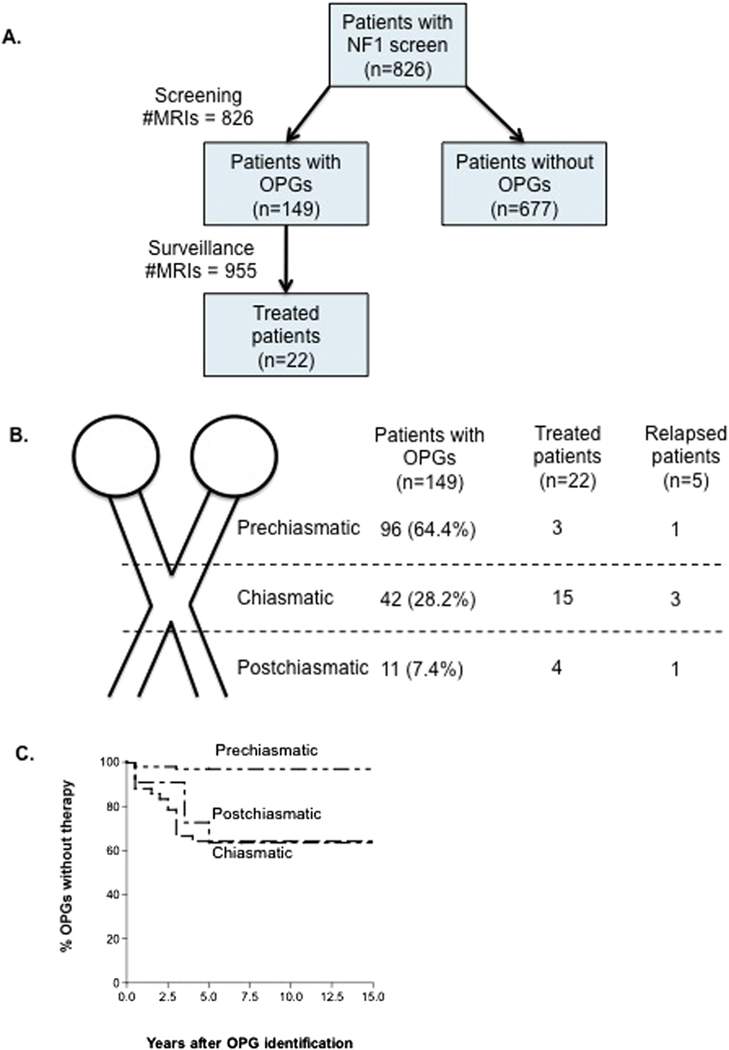
Results: During the 20-year study period, 826 individuals with NF1 (402 females, 424 males) ages 1-9 years were screened for optic pathway gliomas (OPGs) using brain/orbital MRI; 18% were identified with OPGs with a median age at detection of 3 years. Fifteen percent of patients with OPGs had radiologic or clinical progression requiring therapy. Children with chiasmatic and postchiasmatic tumors were more likely to require therapy compared with patients with prechiasmatic OPGs (P < .0001). Patients with visual deficits at the time of diagnosis were more likely to experience visual decline despite therapy when compared with patients treated based on radiologic progression (P < .012).
Conclusions: Our findings confirm that chiasmatic and postchiasmatic OPG in children with NF1 have the highest risk for progression and vision loss. Early identification of OPG by screening MRI prior to the development of vision loss may lead to improved visual outcomes. Children with negative brain and orbital MRI screening at age 15 months or later did not develop symptomatic OPGs.
Copyright © 2015 Elsevier Inc. All rights reserved.
Conflict of interest statement
The authors declare no conflicts of interest.
Figures
References
-
- Sharif S, Upadhyaya M, Ferner R, Majounie E, Shenton A, Baser M, et al. A molecular analysis of individuals with neurofibromatosis type 1 (NF1) and optic pathway gliomas (OPGs), and an assessment of genotype-phenotype correlations. J Med Genet 2011;48:256–60. - PubMed
-
- Moharir M, London K, Howman-Giles R, North K. Utility of positron emission tomography for tumour surveillance in children with neurofibromatosis type 1. Eur J Nucl Med Mol Imaging 2010;37:1309–17. - PubMed
-
- Binning MJ, Liu JK, Kestle JR, Brockmeyer DL, Walker ML. Optic pathway gliomas: a review. Neurosurg Focus 2007;23:E2. - PubMed
-
- Jahraus CD, Tarbell NJ. Optic pathway gliomas. Pediatr Blood Cancer 2006;46:586–96. - PubMed
-
- Nicolin G, Parkin P, Mabbott D, Hargrave D, Bartels U, Tabori U, et al. Natural history and outcome of optic pathway gliomas in children. Pediatr Blood Cancer 2009;53:1231–7. - PubMed
MeSH terms
Grants and funding
LinkOut - more resources
Full Text Sources
Medical
Research Materials
Miscellaneous
