Review
. 2015 Dec:6:183-197.
doi: 10.1016/j.redox.2015.07.008. Epub 2015 Jul 21.

Antioxidant responses and cellular adjustments to oxidative stress

Affiliations
Review

Antioxidant responses and cellular adjustments to oxidative stress

Cristina Espinosa-Diez et al. Redox Biol. 2015 Dec.

Abstract

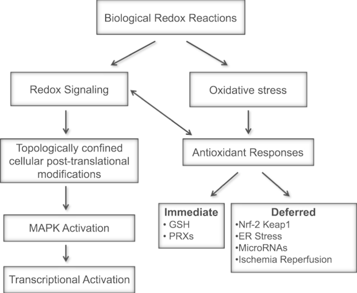
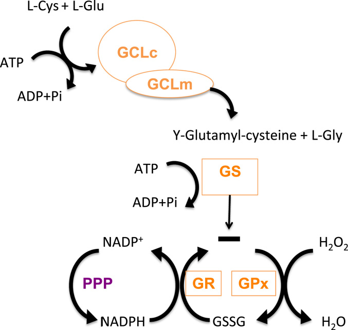
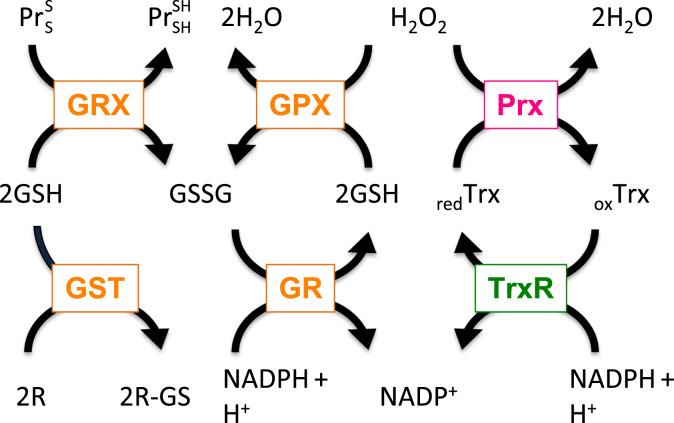
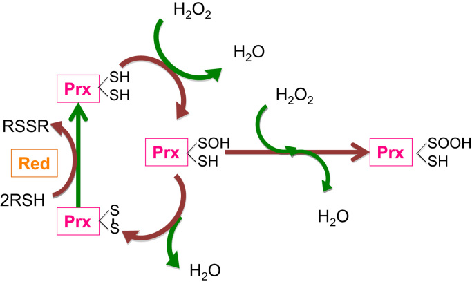
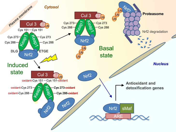
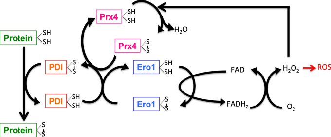
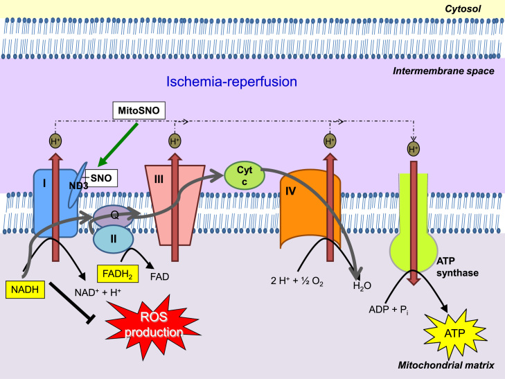
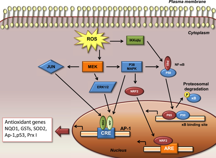
Redox biological reactions are now accepted to bear the Janus faceted feature of promoting both physiological signaling responses and pathophysiological cues. Endogenous antioxidant molecules participate in both scenarios. This review focuses on the role of crucial cellular nucleophiles, such as glutathione, and their capacity to interact with oxidants and to establish networks with other critical enzymes such as peroxiredoxins. We discuss the importance of the Nrf2-Keap1 pathway as an example of a transcriptional antioxidant response and we summarize transcriptional routes related to redox activation. As examples of pathophysiological cellular and tissular settings where antioxidant responses are major players we highlight endoplasmic reticulum stress and ischemia reperfusion. Topologically confined redox-mediated post-translational modifications of thiols are considered important molecular mechanisms mediating many antioxidant responses, whereas redox-sensitive microRNAs have emerged as key players in the posttranscriptional regulation of redox-mediated gene expression. Understanding such mechanisms may provide the basis for antioxidant-based therapeutic interventions in redox-related diseases.

Keywords: Antioxidants; ER stress; Ischemia–reperfusion; Redox signaling; Transcription factors.


Figures

None
Graphical abstract
Fig. 1
Fig. 1
GSH biosynthetic route and GSH cycle. GSH biosynthesis occurs in two different and ATP-dependen steps. The first and limiting step is carried out by GCL, formed by two subunits GCLc and GCLm. In this step l-Glu and l-Cys react to form γ-glutamyl-cisteine. GS is responsible of the second step, that joins l-Gly forming γ-glutamyl-cisteinyl-glicine (GSH). As well as aerobic respiration or other ROS sources increase H2O2, that should be metabolized, in this case GPx generating GSSG. This GSSG could be reduced to GSH again with the help of GR GR, creating a redox cycle, using as reducing agent NAPDH, from Penthoses Phosphate Pathway (PPP).
Fig. 2
Fig. 2
Interconnection of glutaredoxin, peroxiredoxin, thioredoxin, and glutathione containing antioxidant systems. Hydrogen peroxide (H2O2) can be reduced by peroxiredoxins (Prx) or glutathione peroxidases (GPX), which couple reduction of H2O2 with oxidation of glutathione (GSH). Oxidized Prx can be reduced by thioredoxins (Trx). Subsequently, oxidized Trx become reduced by thioredoxin reductase (TrxR) in a NADPH-dependent manner. Oxidized glutathione disulfide (GSSG) is reduced by glutathione reductase (GR) in the presense of NADPH. Further, glutaredoxins (Grx) can reduce disulfide (S–S) bonds in proteins (Pr), and glutathione S transferase (GST) is using GSH to conjugate and thus to detoxify reactive electrophilic compounds (R).
Fig. 3
Fig. 3
Two-step mechanism reaction of a peroxiredoxin dimer with hydrogen peroxide. (i) the sulfhydryl group at the peroxidatic cysteine of one peroxiredoxin (Prx) subunit is oxidized to sulfenic acid (–SOH); (ii) the sulfenic acid condenses with the –SH group at the resolving cysteine from the other subunit to form an intersubunit disulfide bond. The disulfide bond can be reduced by thioredoxin or another reductase (red) depending on the species. Continuous peroxide signaling leads to irreversible hyperoxidation and formation of sulfinic acid (–SOOH) at the peroxidatic cysteine to (shown as the “Hyperoxidation”). The species peroxynitrite (ONOO–) has been included in the cartoon because it is susceptible of reduction by peroxiredoxins.
Fig. 4
Fig. 4
Keap1 dependent regulation of Nrf2. Under basal conditions, Nrf2 is sequestered in the cytosol by a Keap1 homodimer, which facilitates the ubiquitination and proteasomal degradation of Nrf2. Inducers react with specific cysteine residues in Keap1, leading to the release of Nrf2 and allowing its nuclear translocation. In the nucleus, Nrf2 heterodimerizes with small Maf proteins and binds to the antioxidant response element (ARE), activating the expression of a battery of cytoprotective genes.
Fig. 5
Fig. 5
Protein folding in the endoplasmic reticulum is coupled to ROS formation. The ER enzymes PDI and ERO1 are crucial for protein folding. PDI oxidizes thiol (SH) groups, i.e. generates disulfide bonds, in the folding substrates, thereby it accepts electrons (e) and becomes reduced. Ero1 re-oxidizes PDI by using its FAD moiety to transfer electrons from PDI to molecular oxygen (O2) to form hydrogen peroxide (H2O2), which may give rise to further formation of ROS. Hydrogen peroxide may also be used by peroxiredoxin 4 (Prx4) to oxidize PDI, thereby increasing the efficiency of Ero1-dependent disulfide bond formation.
Fig. 6
Fig. 6
S-Nitrosylation of ND3 Cys39 protects from IR injury. The low activity of complex I after ischemia causes the ND3 Cys39 to become exposed. Reperfusion of ischemic tissue rapidly reactivates complex I and generates superoxide leading to oxidative damage and cell death. The presence of MitoSNO or other S-nitrosylating agents at reperfusion, causes the exposed ND3 cysteine to become S-nitrosylated, holding complex I in a low activity state and decreasing ROS production. Complex I is gradually reactivated through the reduction of the S-nitrosothiol by glutathione and thioredoxin.
Fig. 7
Fig. 7
Induction of antioxidant responses by ROS-mediated activation of cytosolic cell signaling pathways. ROS drive activation of MAPKs. ERK and JNK are involved in recruiting c-Fos and c-Jun to the nucleus where they activate the transcription factor AP-1, whereas activation of p38 and IKK is important in the transcriptional activation of NF-ĸB. Both of these factors are important in regulating diverse genes, including antioxidants (p53, NQO1, GSTs, SOD2, Ap-1,p53, and Prx I).

References

    1. Hwang C., Sinskey A.J., Lodish H.F. Oxidized redox state of glutathione in the endoplasmic reticulum. Science. 1992;257(5076):1496–1502. - PubMed
    1. Pastore A., Piemonte F. S-glutathionylation signaling in cell biology: progress and prospects. Eur. J. Pharm. Sci. 2012;46(5):279–292. - PubMed
    1. Ray P.D., Huang B.W., Tsuji Y. Reactive oxygen species (ROS) homeostasis and redox regulation in cellular signaling. Cell Signal. 2012;24(5):981–990. - PMC - PubMed
    1. Rahman I. Glutathione, stress responses, and redox signaling in lung inflammation. Antioxid. Redox Signal. 2005;7(1–2):42–59. - PubMed
    1. Haddad J.J., Harb H.L. L-gamma-glutamyl-l-cysteinyl-glycine (glutathione; GSH) and GSH-related enzymes in the regulation of pro- and anti-inflammatory cytokines: a signaling transcriptional scenario for redox(y) immunologic sensor(s)? Mol. Immunol. 2005;42(9):987–1014. - PubMed

Publication types

MeSH terms