. 2015 Dec;17(12):5073-88.
doi: 10.1111/1462-2920.12991. Epub 2015 Sep 10.

Vertical stratification of matrix production is essential for physical integrity and architecture of macrocolony biofilms of Escherichia coli

Affiliations

Vertical stratification of matrix production is essential for physical integrity and architecture of macrocolony biofilms of Escherichia coli

Diego O Serra et al. Environ Microbiol. 2015 Dec.

Abstract

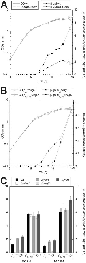
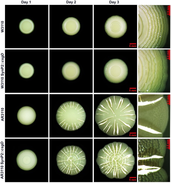
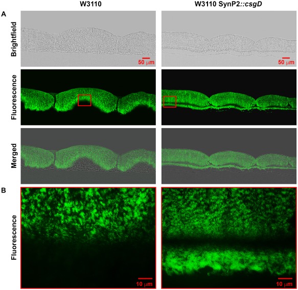
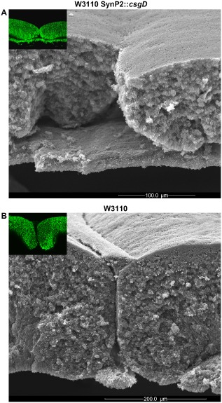
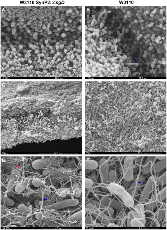
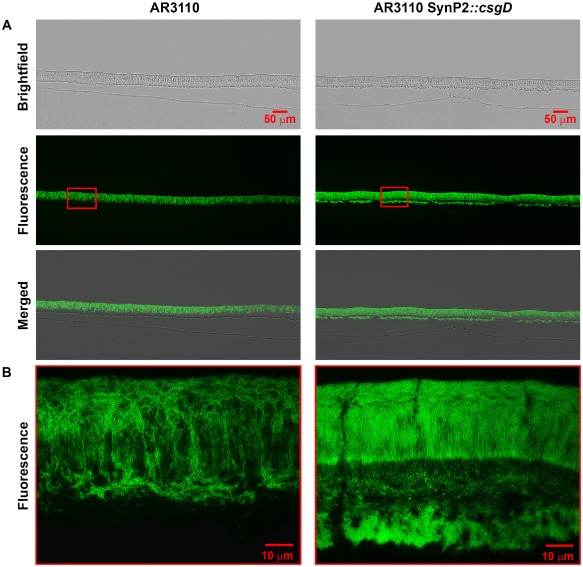
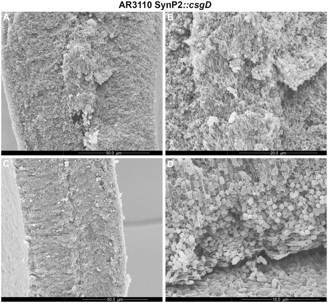
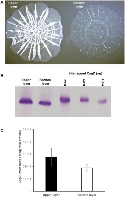
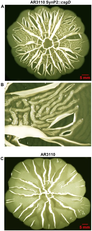
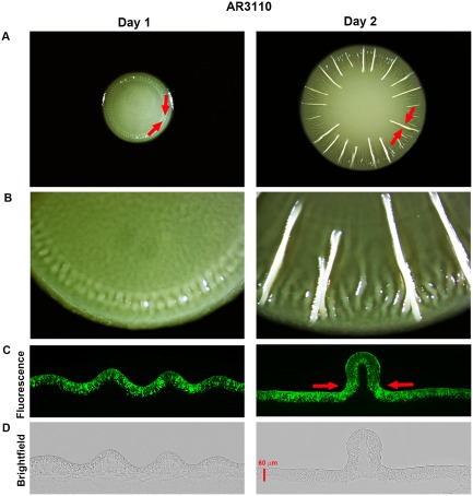
Bacterial macrocolony biofilms grow into intricate three-dimensional structures that depend on self-produced extracellular polymers conferring protection, cohesion and elasticity to the biofilm. In Escherichia coli, synthesis of this matrix - consisting of amyloid curli fibres and cellulose - requires CsgD, a transcription factor regulated by the stationary phase sigma factor RpoS, and occurs in the nutrient-deprived cells of the upper layer of macrocolonies. Is this asymmetric matrix distribution functionally important or is it just a fortuitous by-product of an unavoidable nutrient gradient? In order to address this question, the RpoS-dependent csgD promoter was replaced by a vegetative promoter. This re-wiring of csgD led to CsgD and matrix production in both strata of macrocolonies, with the lower layer transforming into a rigid 'base plate' of growing yet curli-connected cells. As a result, the two strata broke apart followed by desiccation and exfoliation of the top layer. By contrast, matrix-free cells at the bottom of wild-type macrocolonies maintain colony contact with the humid agar support by flexibly filling the space that opens up under buckling areas of the macrocolony. Precisely regulated stratification in matrix-free and matrix-producing cell layers is thus essential for the physical integrity and architecture of E. coli macrocolony biofilms.


Figures

Figure 1
Figure 1
Comparison of the expression of curli genes under the control of CsgD expressed either from its natural promoter (P wt) or a synthetic vegetative standard promoter (PSynP2). Derivatives of E . coliK‐12 strains W3110 (A, B, C) and AR3110 (C) carrying a single‐copy csgB::lacZ reporter fusion as well as the mutations indicated directly on the figures were grown in LB medium at 28°C. Optical density at 578 nm (OD 578) was monitored (open symbols in A and B) and specific β‐galactosidase activities were determined. In (A) the csgB::lacZ expression pattern along the growth cycle and RpoS dependence was assayed in W3110 expressing CsgD under Synp2 control. In (B) the timing of induction of csgB::lacZ (expression normalized to 1.0) was compared in strains that express CsgD either under natural or Synp2 control. (C) Expression of CsgD under SynP2 control eliminates c‐di‐GMP dependence of csgB::lacZ, as tested using knockout mutations in the genes encoding the DGCs and PDEs that make up the central c‐di‐GMP switch that controls csg D in wild‐type E . coliK‐12 (Lindenberg et al., 2013).
Figure 2
Figure 2
Macrocolony morphology is altered by expression of CsgD under the control of the SynP2 promoter. Time‐course of macrocolony formation in TS‐containing salt‐free LB plates of strains W3110, AR3110 and respective derivatives expressing CsgD under the control of the SynP2 promoter. Right panels show enlarged view of the respective macrocolonies at day 3.
Figure 3
Figure 3
SynP2‐dependent CsgD expression results in production of a curli fibre matrix in the upper and lower layers of macrocolonies of strain W3110. Macrocolony biofilms of strain W3110 expressing CsgD either from its natural promoter or the SynP2 promoter were grown for 3 days on salt‐free LB medium supplemented with TS, then cryo‐embedded and sectioned perpendicular to the plane of the macrocolony at a thickness of 5 μm. A. Macrocolony sections were visualized at low magnification with brightfield (top panels) and fluorescence (middle panels) and shown as merged images (bottom panels). B. Enlarged views of TS fluorescence patterns in macrocolony areas boxed in red in panel A.
Figure 4
Figure 4
SynP2‐driven CsgD expression transforms the lower layer of growing cells in a W3110 macrocolony into a rigid ‘base plate’. Side‐view SEM images at low magnification showing a cross section of macrocolonies of strain W3110 expressing CsgD either from SynP2 (A) or its natural promoter (B). The upper‐left insets are low‐magnification fluorescence images of corresponding macrocolony regions showing the TS‐stained curli fibre matrix. A false‐coloured version of a section of the SEM image shown in (B) has previously been published (Serra and Hengge, 2014).
Figure 5
Figure 5
SynP2‐driven CsgD expression results in curli production by cells in all zones of macrocolonies of strain W3110. High‐resolution SEM images of macrocolonies of strain W3110 expressing CsgD either from its natural promoter (right panels) or the SynP2 promoter (left panels). SEM images offer detailed views of the macrocolony surface at the breaks between the rings (A and B) and the macrocolony interior at the lower layer (C and D). A blue arrow in panel B points to elongated dividing cells that are free of curli. (E) Magnified SEM view of the lower layer of W3110 SynP2::csg D macrocolony showing elongated flagellated cells surrounded by a dense network of curli fibres. (F) Magnified SEM view of the lower layer of W3110 macrocolony showing elongated cells interconnected by flagella. Blue arrows in panels E and F point to flagella filaments. A red arrow in panel E indicates the network of curli fibres.
Figure 6
Figure 6
SynP2‐dependent CsgD expression occurs in both physiological layers and has a pronounced effect on supracellular architecture of macrocolonies of strain AR3110. Macrocolony biofilms of strain AR3110 expressing CsgD either from its natural promoter or the SynP2 promoter were grown for 3 days on salt‐free LB medium supplemented with TS, then cryo‐embedded and sectioned perpendicular to the plane of the macrocolony at a thickness of 5 μm. A. Macrocolony sections were visualized at low magnification with brightfield (top panels) and fluorescence (middle panels) and shown as merged images (bottom panels). B. Enlarged views of TS fluorescence pattern in macrocolony areas boxed in red in panel A.
Figure 7
Figure 7
High‐resolution SEM analysis of altered supracellular architecture of macrocolonies of strain AR3110 expressing CsgD under SynP2 control. SEM images of representative cross‐sections at the tip (A) and middle body (C) of a macrocolony ridge of strain AR3110 SynP2 ::csg D. (B) Magnified SEM view of the central area of the ridge tip presented in A, offering details of a chunk of rod‐shaped cells tightly encased by matrix. (D) Magnified SEM view of the zone of transition between the lower and upper layers of an AR3110 SynP2::csg D macrocolony. The image offers detailed view of the spatial arrangement of cells and sheet‐like envelopes – distinctive of cellulose – in the lower part of the upper macrocolony layer.
Figure 8
Figure 8
Physical separation of the integral upper and lower layers of macrocolonies of strain AR3110 expressing CsgD under SynP2 control. Three‐day‐old AR3110 SynP2::csg D macrocolonies grown on agar plates were gently overlaid with liquid medium, which results in the upper layer detaching and floating away. (A) The two macrocolony layers with the upper layer floating and the lower layer attached to the agar surface. (B) Immunoblot showing CsgD protein in the two layers, with purified His6‐tagged CsgD used for the quantification shown in (C).
Figure 9
Figure 9
Long‐term desiccation and exfoliation of the upper layer of macrocolonies of strain AR3110 expressing CsgD under SynP2 control. A. Top view image of an AR3110 SynP2::csg D macrocolony grown on agar plates for 10 days. The image shows severe disruption of the upper macrocolony layer with multiple irregular breakages, some of which give rise to an exfoliation pattern. B. Magnified view of a region of the AR3110 SynP2::csg D macrocolony presented in A, offering details of breakages and exfoliated sectors of the upper macrocolony layer. C. Top view image of a macrocolony biofilm of strain AR3110 grown on agar plates for 10 days.
Figure 10
Figure 10
Matrix‐free cells of the lower layer maintain tight contact of the colony to the agar surface when young AR3110 macrocolonies buckle up into ridges. (A) Images of a representative AR3110 macrocolony after 1 and 2 days of growth on TS‐containing salt‐free LB plates. Red arrows indicate areas under particular mechanical tension generated by the increasingly constrained space that cells already connected by the elastic curli‐cellulose matrix can enter (Serra et al., 2013a). (B) Enlarged views of the respective macrocolonies offering details of emerging ridges. (C) Fluorescence and (D) brightfield images of vertical cross‐sections of corresponding macrocolonies presented in A. Red arrows in D represent convergent mechanical forces that promote colony buckling up.

References

    1. Aguilar, C. , Vlamakis, H. , Losick, R. , and Kolter, R. (2007) Thinking about Bacillus subtilis as a multicellular organism. Curr Opin Microbiol 10: 638–643. - PMC - PubMed
    1. Allesen‐Holm, M. , Barken, K.B. , Yang, L. , Klausen, M. , Webb, J.S. , Kjelleberg, S. , et al (2006) A characterization of DNA release in Pseudomonas aeruginosa cultures and biofilms. Mol Microbiol 59: 1114–1128. - PubMed
    1. Asally, M. , Kittisopikul, M. , Rué, P. , Du, Y. , Hu, Z. , Cagatay, T. , et al (2012) Localized cell death focuses mechanical forces during 3D patterning in a biofilm. Proc Natl Acad Sci U S A 109: 18891–18896. - PMC - PubMed
    1. Branda, S.S. , Vik, A. , Friedman, L. , and Kolter, R. (2005) Biofilms: the matrix revisited. Trends Microbiol 13: 20–26. - PubMed
    1. Cerda, E. , and Mahadevan, L. (2003) Geometry and physics of wrinkling. Phys Rev Lett 90: 074302. - PubMed

Publication types

MeSH terms