. 2015 Oct;24(10):1671-85.
doi: 10.1002/pro.2762. Epub 2015 Aug 17.

Transient sampling of aggregation-prone conformations causes pathogenic instability of a parkinsonian mutant of DJ-1 at physiological temperature

Affiliations

Transient sampling of aggregation-prone conformations causes pathogenic instability of a parkinsonian mutant of DJ-1 at physiological temperature

Nicole M Milkovic et al. Protein Sci. 2015 Oct.

Abstract

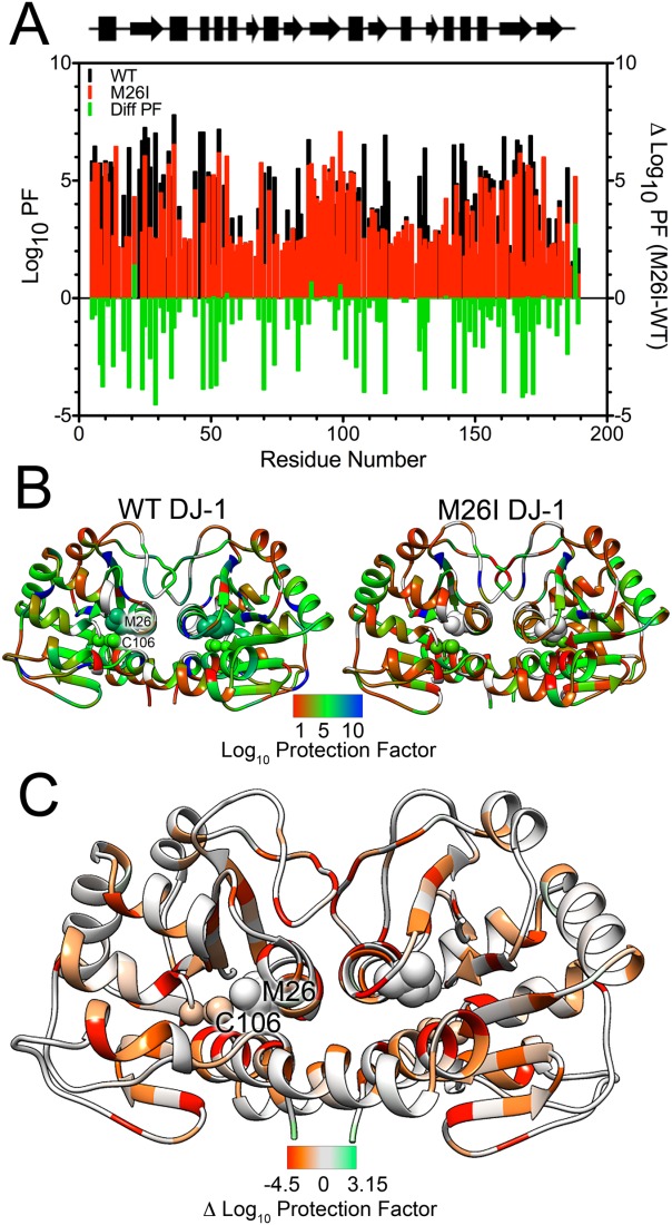
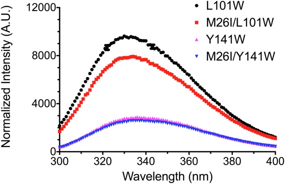
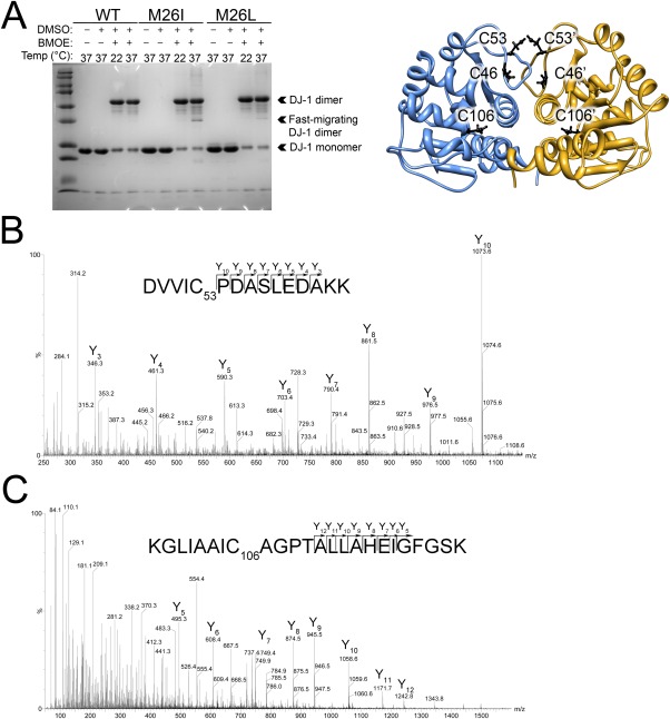
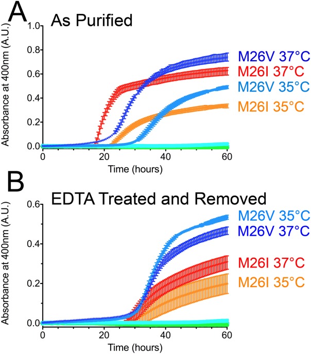
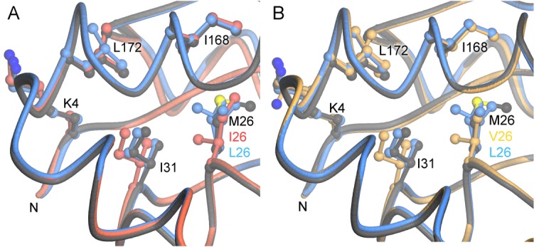
Various missense mutations in the cytoprotective protein DJ-1 cause rare forms of inherited parkinsonism. One mutation, M26I, diminishes DJ-1 protein levels in the cell but does not result in large changes in the three-dimensional structure or thermal stability of the protein. Therefore, the molecular defect that results in loss of M26I DJ-1 protective function is unclear. Using NMR spectroscopy near physiological temperature, we found that the picosecond-nanosecond dynamics of wild-type and M26I DJ-1 are similar. In contrast, elevated amide hydrogen/deuterium exchange rates indicate that M26I DJ-1 is more flexible than the wild-type protein on longer timescales and that hydrophobic regions of M26I DJ-1 are transiently exposed to solvent. Tryptophan fluorescence spectroscopy and thiol crosslinking analyzed by mass spectrometry also demonstrate that M26I DJ-1 samples conformations that differ from the wild-type protein at 37°C. These transiently sampled conformations are unstable and cause M26I DJ-1 to aggregate in vitro at physiological temperature but not at lower temperatures. M26I DJ-1 aggregation is correlated with pathogenicity, as the structurally similar but non-pathogenic M26L mutation does not aggregate at 37°C. The onset of dynamically driven M26I DJ-1 instability at physiological temperature resolves conflicting literature reports about the behavior of this disease-associated mutant and illustrates the pitfalls of characterizing proteins exclusively at room temperature or below, as key aspects of their behavior may not be apparent.

Keywords: DJ-1; PARK7; Parkinson's disease; conformational dynamics; protein stability; thiol crosslinking.


Figures

Figure 1
Figure 1
Wild-type and M26I DJ-1 have similar ps-ns dynamics at 35°C. The general order parameter (S2) for wild-type (WT, black) and M26I DJ-1 (red) at 35°C obtained from FAST ModelFree are plotted per residue. Picosecond-nanosecond (ps-ns) motions for both proteins are nearly identical at 35°C. The regions without a reported value indicate amino acids that could not be analyzed by FAST ModelFree, primarily due to unobserved or overlapping resonances in the NOE spectra. A schematic of the secondary structure of DJ-1 is shown below for reference (rectangles=β-sheet, arrows=α-helix).
Figure 2
Figure 2
HDX-detected dynamics are globally enhanced in M26I DJ-1 at 35°C. A: Calculated log10 HDX protection factors (Log10PF, left Y-axis) for wild-type (WT, black) and M26I (red) DJ-1. Lower Log10PF values correspond to residues that more rapidly exchange backbone amide hydrogen atoms with solvent deuterons. Difference Log10PF values are shown in the green bars (right Y-axis) and quantify the lower protection (more rapid exchange) of M26I DJ-1 compared to wild-type protein. B: Log10PF values from (A) have been mapped onto the structure of wild-type (WT, left) and M26I (right) DJ-1 dimer. Met26 and Cys106 are shown in sphere rendering and labeled in one monomer. The apparent proximity of these residues is an effect of this view; they are approximately 18 Å apart in each monomer. All residues for which a Log10PF could not be assigned are shown in gray. Red indicates lower protection (faster exchange) with blue indicates greater protection (slower exchange). M26I DJ-1 is more exchange-active than WT DJ-1, particularly in the hydrophobic core of the protein. C: Difference Log10PF (M26I Log10PF – WT Log10PF) mapped on to the dimer of M26I DJ-1. Red indicates areas that more rapidly exchange with solvent in M26I DJ-1 than in wild-type protein and green indicates the converse. Areas that are similar between the two proteins are gray. The enhanced exposure of the hydrophobic core of M26I DJ-1 to solvent is evident.
Figure 3
Figure 3
Tryptophan fluorescence emission spectra reveal site-specific differences in M26I DJ-1 solvent exposure. Tryptophan emission spectra for L101W (black circle), M26I/L101W (red square), Y141W (magenta triangle), and M26I/Y141W (blue inverted triangle) obtained at 37°C. The M26I mutation results in quenching of L101W DJ-1 fluorescence, indicating Trp101 is in an altered environment in M26I DJ-1. Leu101 is in a dynamic area of DJ-1 that is sensitive to the M26I substitution in HDX (Log10PF WTL101=4.35, Log10PF M26IL101=1.75.) In contrast, no differences are observed in the spectra for Y141W and M26I/Y141W DJ-1. Tyr141 is in a dynamic region of DJ-1 that is not sensitive to the M26I mutation (Log10PF WTY141=2.09, Log10PF M26IY141=2.08.).
Figure 4
Figure 4
Thiol crosslinking shows that M26I DJ-1 is more dynamic than wild-type DJ-1 at physiological temperature. A: Left: Cysteine crosslinking with BMOE for wild-type (WT), M26I, and an engineered M26L mutant DJ-1 at 22 and 37°C resolved by SDS-PAGE. At 37°C, M26I DJ-1 forms a faster migrating crosslinked dimeric species (indicated) that is absent at 22°C and also not seen for WT DJ-1. This faster-migrating species is present but ∼6-fold less abundant in the M26L mutant that relieves the steric conflict introduced by the M26I mutation. Monomer and dimer DJ-1 bands are indicated. Right: Location of all six cysteine residues in the DJ-1 dimer is shown as a ribbon diagram. B: The MS/MS spectrum of the Cys53-Cys53 BMOE crosslinked peptide in the dominant dimer band (“DJ-1 dimer” in A). The sequence and predicted Y daughter ion series are shown with all detected Y ion species labeled in the MS/MS spectrum. C: The MS/MS spectrum of the Cys106-Cys106 BMOE crosslinked peptide from M26I DJ-1, which is only found in the fast-migrating dimeric species at 37°C (“Fast-migrating DJ-1 dimer” in A). As in (B), the sequence and predicted Y daughter ion series are shown with Y ion peaks labeled.
Figure 5
Figure 5
M26I DJ-1 is aggregation-prone at physiological temperature in vitro. A: Aggregation of wild-type (WT), M26I, M26L, and M26V at 30, 35, and 37°C. There is no observable aggregation for wild-type and M26L DJ-1 at any of these temperatures (cyan and green symbols at baseline). In contrast, M26I and M26V DJ-1 aggregate extensively at 35 and 37°C (labeled) but not at 30°C. B: Aggregation was performed as in (A), but the samples were treated with 10 mM EDTA and dialyzed to remove any trace metal contamination from the proteins before the start of the experiment. M26I and M26V DJ-1 still aggregate at 35 and 37°C (labeled), although to a lesser extent than in (A). All samples were measured in triplicate (plotted as avg±SD.).
Figure 6
Figure 6
The aggregation-inducing M26I and M26V mutations cause similar steric conflicts with Ile31. A: The local environment of residue 26 is shown for wild-type (dark gray), M26L (blue), and M26I (red) DJ-1. B: A similar superposition to (A), but with M26V (gold). The conflict with Ile31 is the most prominent structural change resulting from mutation of Met26 to Ile (A) or Val (B). The M26L mutation alleviates this steric clash. This steric conflict could communicate changes at the buried residue 26 to the exterior of the protein, particularly the region containing both the N-terminus and C-proximal residues such as Ile168 and Leu172 (labeled). This figure was made with POVScript+.

References

    1. Bonifati V, Rizzu P, van Baren MJ, Schaap O, Breedveld GJ, Krieger E, Dekker MC, Squitieri F, Ibanez P, Joosse M, van Dongen JW, Vanacore N, van Swieten JC, Brice A, Meco G, van Duijn CM, Oostra BA, Heutink P. Mutations in the DJ-1 gene associated with autosomal recessive early-onset parkinsonism. Science. 2003;299:256–259. - PubMed
    1. Greenamyre JT, Hastings TG. Biomedicine. Parkinson’s—divergent causes, convergent mechanisms. Science. 2004;304:1120–1122. - PubMed
    1. Waak J, Weber SS, Gorner K, Schall C, Ichijo H, Stehle T, Kahle PJ. Oxidizable residues mediating protein stability and cytoprotective interaction of DJ-1 with apoptosis signal-regulating kinase 1. J Biol Chem. 2009;284:14245–14257. - PMC - PubMed
    1. Jin J, Li GJ, Davis J, Zhu D, Wang Y, Pan C, Zhang J. Identification of novel proteins associated with both alpha-synuclein and DJ-1. Mol Cell Proteomics. 2007;6:845–859. - PubMed
    1. Clements CM, McNally RS, Conti BJ, Mak TW, Ting JP. DJ-1, a cancer- and Parkinson’s disease-associated protein, stabilizes the antioxidant transcriptional master regulator Nrf2. Proc Natl Acad Sci USA. 2006;103:15091–15096. - PMC - PubMed

Publication types

Substances