. 2016 Jan 15;138(2):417-27.
doi: 10.1002/ijc.29709. Epub 2015 Aug 17.

Mesenchymal stem cells regulate melanoma cancer cells extravasation to bone and liver at their perivascular niche

Affiliations

Mesenchymal stem cells regulate melanoma cancer cells extravasation to bone and liver at their perivascular niche

Diego Correa et al. Int J Cancer. .

Abstract

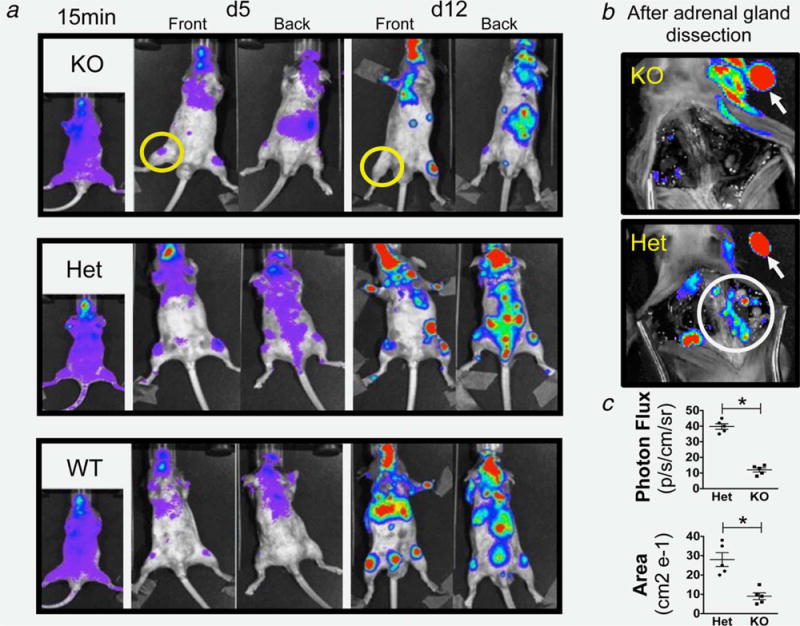
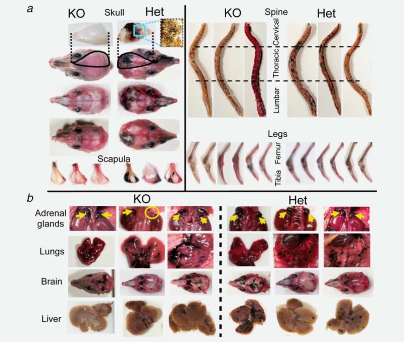
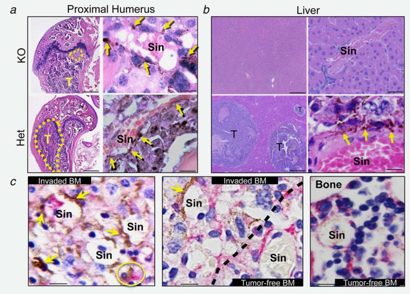
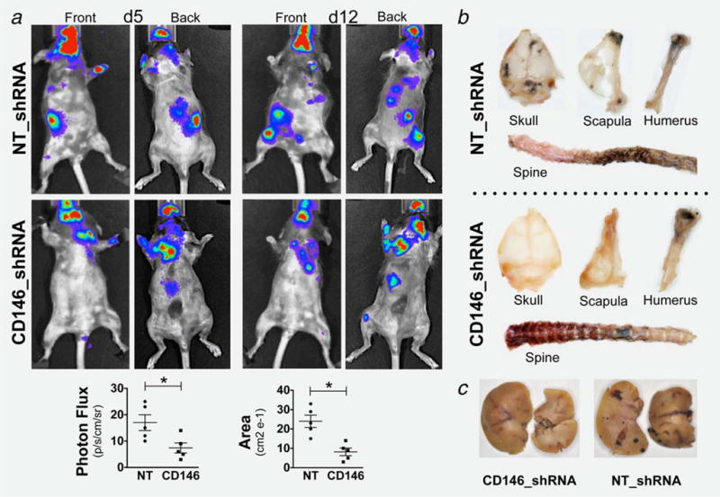
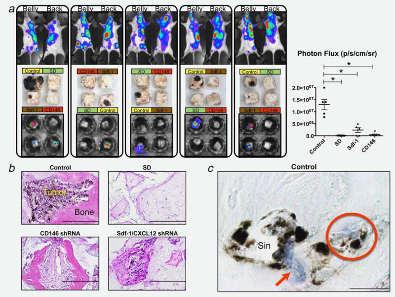
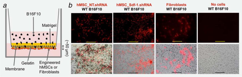
Skeleton and liver are preferred organs for cancer dissemination in metastatic melanoma negatively impacting quality of life, therapeutic success and overall survival rates. At the target organ, the local microenvironment and cell-to-cell interactions between invading and resident stromal cells constitute critical components during the establishment and progression of metastasis. Mesenchymal stem cells (MSCs) possess, in addition to their cell progenitor function, a secretory capacity based on cooperativity with other cell types in injury sites including primary tumors (PT). However, their role at the target organ microenvironment during cancer dissemination is not known. We report that local MSCs, acting as pericytes, regulate the extravasation of melanoma cancer cells (MCC) specifically to murine bone marrow (BM) and liver. Intra-arterially injected wild-type MCC fail to invade those selective organs in a genetic model of perturbed pericyte coverage of the vasculature (PDGF-B(ret/ret)), similar to CD146-deficient MCC injected into wild type mice. Invading MCC interact with resident MSCs/pericytes at the perivascular space through co-expressed CD146 and Sdf-1/CXCL12-CXCR4 signaling. Implanted engineered bone structures with MSCs/pericytes deficient of either Sdf-1/CXCL12 or CD146 become resistant to invasion by circulating MCC. Collectively, the presence of MSCs/pericytes surrounding the target organ vasculature is required for efficient melanoma metastasis to BM and liver.

Keywords: CD146; Sdf-1; mesenchymal stem cells (MSCs); metastatic melanoma; pericytes.


Figures

Figure 1
Figure 1
BLI of injected PDGF-B mutant and control mice: (a) Imaging 15 min, 5 and 12 days postinjection, showing increased skeletal invasion (limbs and spine) in WT and Het mice compared to PDGF-B mutant mice and the disappearance of a metastatic signal at 12 days in PDGF-B mutant mice (yellow circles). (b) Absence in PDGF-B mutant and persistence in Het mice of the spine signal (white circle) after adrenal gland removal (white arrows) confirming the nonspinal origin of the signal in PDGF-B mutant spine (yellow arrow in A). (c) Quantification of signal (photon flux and area covered by tumors) showing statistical difference between PDGF-B mutant and Het mice (* = p < 0.01). Data are represented as mean ± SEM. Representative mice of n = 15 (5 per group).
Figure 2
Figure 2
Gross inspection of distant melanoma dissemination: (a) Craniofacial and appendicular invasion by MCC exhibiting a significant reduction in PDGF-B mutant mice. All skulls and scapulae in the mutants were clear of metastases or had only few/small foci, which contrasts sharply with the multiple major/multifocal invasion observed in Het mice. Spines in PDGF-B mutant mice were clear of metastasis in 2/5 mice, or harbored only 1–2 small foci restricted to one vertebral segment in the remaining three animals. WT and Het mice had multiple multisegment metastases in all animals. Both distal femur and proximal tibia were compromised bilaterally in all WT and Het controls, while bilateral invasion was observed in only one of the five PDGF-B mutant mice, with remaining four mice exhibiting only one compromised leg that was restricted to the proximal tibia. (b) Melanoma invasion to other target organs with comparable results in both genotypes except for liver (reduced in PDGF-B mutant mice). Yellow arrows: adrenal glands. Yellow circle: adrenal gland agenesis. Three representative mice shown of n = 15 (5 per group).
Figure 3
Figure 3
Histology of metastatic bone tumors. (a, b) Metastatic tumors (T) in distal femur (a) and liver (b) are smaller or absent in PDGF-B mutant mice. Bars = 200 μm (low magnification) and 10 μm (high magnification). Melanin-producing B16F10 cells (yellow arrows) localize in the abluminal side of BM sinusoids (Sin), extending to the tissue parenchyma (impaired in PDGF-B mutant mice). (c) CD146 IHC in BM sections from Het mice. Engrafted B16F10 cells (yellow arrows) physically associate with CD146-positive BMMSC/pericytes (pink signal) at the perivascular space and inside the parenchyma (yellow circle). Dotted line = boundary between tumor-invaded and tumor-free BM. Bar = 10 μm.
Figure 4
Figure 4
Invasion of engineered B16F10 MCC: Reduced invasion of CD146-silenced MCC to craniofacial, appendicular structures and liver (compared to NT-shRNA), evaluated by BLI (a) and gross examination (b and c). BLI signal reached statistical significance (* = p < 0.01). Data are represented as mean ± SEM. Representative mice of n = 6 (3 per group).
Figure 5
Figure 5
B16F10 MCC invasion to humanized extraskeletal ossicles. (a) MCC invasion of the skeleton (top row BLI) and specific implanted ossicles, evaluated by direct examination after harvesting (middle row) and by BLI (bottom row). The signal from the cubes BLI was quantified (photon flux) giving statistical difference of all groups compared to control (* = p < 0.01). Data are represented as mean ± SEM. Representative mice and cubes of n = 8. (b) Histological analysis (H&E staining) of harvested ossicles shows significant melanoma invasion in structures made with control MSC while significantly reduced or absent with Sdf-1 and CD146-silenced cells. SD cubes exhibit no bone and vasculature formation. Bar = 200 μm. (c) Immunolocalization of CD146 (blue signalred arrow) in sections from Control ossicles showing invading MCC physically associated with MSC/pericytes at the perivascular space surrounding sinusoids (Sin), and advancing towards the tissue parenchyma as a cell complex (red circle). Bar = 10 μm.
Figure 6
Figure 6
In vitro transendothelial migration assay: (a) Schematic representation of the modified TEM. (b–top row) Fluorescence microscopy of DiI-labeled (red) hMSC expressing either nontarget (NT) or Sdf-1 silencing vectors (Sdf-1_shRNA) and papillary dermal Fibroblasts seeded at the bottom surface of an 8 μm pore diameter insert membrane. (b–bottom row) Merged bright field and fluorescence microscopy showing B16F10 melanoma cell invasion to the membrane and interaction with seeded cells (Bar = 200 μm). Representative pictures from three independent experiments.

References

    1. Caplan AI. Mesenchymal stem cells. J Orthop Res. 1991;9:641–650. - PubMed
    1. Caplan AI, Correa D. The MSC: an injury drugstore. Cell Stem Cell. 2011;9:11–15. - PMC - PubMed
    1. Caplan AI. All MSCs are pericytes? Cell Stem Cell. 2008;3:229–230. - PubMed
    1. Crisan M, Yap S, Casteilla L, et al. A perivascular origin for mesenchymal stem cells in multiple human organs. Cell Stem Cell. 2008;3:301–313. - PubMed
    1. da Silva Meirelles L, Caplan AI, Nardi NB. In search of the in vivo identity of mesenchymal stem cells. Stem Cells. 2008;26:2287–2299. - PubMed

Publication types