Comparative Study
. 2016 Mar-Apr;26(2):240-6.
doi: 10.1111/jon.12281. Epub 2015 Aug 3.

Comparison of Glioblastomas and Brain Metastases using Dynamic Contrast-Enhanced Perfusion MRI

Affiliations
Comparative Study

Comparison of Glioblastomas and Brain Metastases using Dynamic Contrast-Enhanced Perfusion MRI

Brian C Jung et al. J Neuroimaging. 2016 Mar-Apr.

Abstract

Purpose: To compare glioblastoma and brain metastases using T1-weighted dynamic contrast-enhanced (DCE)-MRI perfusion technique.

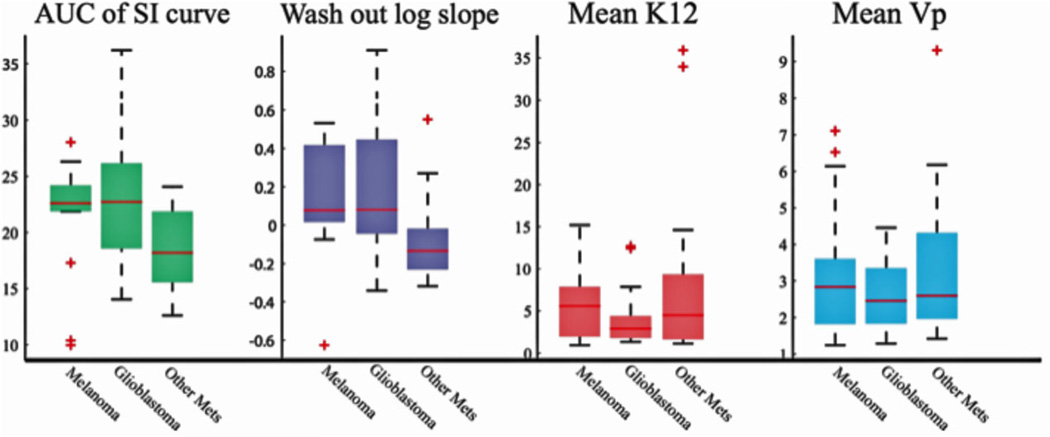
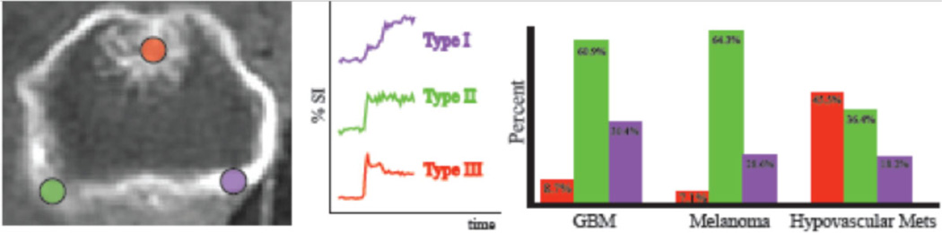
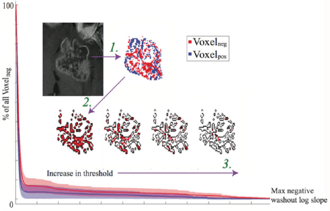
Methods: 26 patients with glioblastoma and 32 patients with metastatic brain lesions with no treatment who underwent DCE-MRI were, retrospectively, analyzed. DCE perfusion parameters K(trans) and Vp were calculated for the whole tumor. Signal intensity time curves were quantified by calculating the area under the curve (AUC) and the logarithmic slope of the washout phase to explore the heterogeneous tumor characteristics.

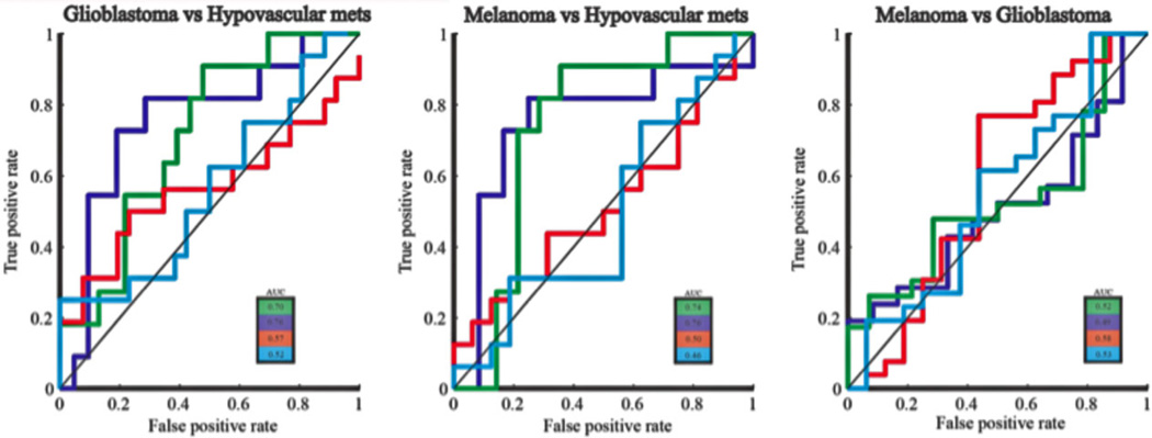
Results: Glioblastoma did not differ from all brain metastases in K(trans) (P = .34) or Vp (P = .47). Glioblastoma and melanoma metastases differed from hypovascular metastases in AUC and log slope of the washout phase of the signal intensity time curve (P < .05); however, glioblastoma and melanoma metastases did not differ from each other (AUC: P = .78, Log slope: P = .77). Glioblastoma and melanoma metastases differed from hypovascular metastases in the ratio of Voxelneg /Voxelpos (P< .03); however, they did not differ from each other. Glioblastoma and melanoma metastases differed from each other in Voxelneg_threshold at higher negative log slope threshold.

Conclusion: DCE-MRI showed that it has a potential to differentiate glioblastomas, melanoma metastases and hypovascular brain tumors. Logarithmic slope of the washout phase and AUC of the signal intensity time curve were shown to be the best discriminator between hypervascular and hypovascular neoplasms.

Keywords: Glioblastoma; brain; dynamic contrast-enhanced (DCE) MRI; metastasis; neoplasm.


Figures

Fig 1
Fig 1
The morphology of the signal intensity time curve could differentiate hypervascular brain tumors from hypovascular brain tumors: Hypervascular brain tumors (glioblastoma and melanoma metastases) differed from hypovascular brain metastases in the AUC and the logarithmic slope of the washout phase of the signal intensity time curve; however, glioblastoma and melanoma metastases could not be differentiated from each other based on the morphological characteristics of the signal intensity time curve. Ktrans and Vp could not differentiate between the three pathologies.
Fig 2
Fig 2
ROC curve shows that AUC and log slope of the washout phase could successfully discriminate between hypervascular neoplasms from hypovascular metastases: ROC analysis showed that the logarithmic slope of the washout phase had the highest discriminatory power in differentiating hypervascular tumors (glioblastoma and melanoma) from hypovascular metastases (AUC = .76), followed by the AUC of the signal intensity time curve (AUC = .70 and .74, respectively). Neither of these two variables could differentiate glioblastoma from metastatic melanoma (AUC ≤ .52).
Fig 3
Fig 3
Perfusion characteristics at the region of maximal enhancement: At the region of maximal enhancement, hypovascular metastases showed an increased number of type III signal intensity time curves, while both types of hypervascular tumors showed greater number of type I and II morphology.
Fig 4
Fig 4
Glioblastoma shows increased number of Voxelneg at higher negative log slope threshold: Logarithmic slope of the washout phase of the signal intensity time curve was calculated for each voxel within the tumor region. Arrow #1: Each voxel was then classified as Voxelneg or Voxelpos based on the value of the logarithmic slope (Voxelneg = [log slope < 0]; Voxelpox = [log slope > 0]). Voxels that showed poor linear correlation in the washout phase were excluded from the analysis. Arrow #2: All the Voxelneg were then selected for further analysis (total number of Voxelneg; threshold = 0). Arrow #3: The Voxelneg was then calculated at different negative slope thresholds (Voxelneg_threshold = [log slope < threshold], Voxelplateau_threshold = [threshold < log slope < 0], Voxelpox_threshold = [log slope > threshold]). The number of Voxelneg_threshold at different threshold (red filled pixels) was then expressed as a fraction of the total number of Voxelneg (black outline). At higher threshold values, glioblastoma showed an increased ratio of (Voxelneg_threshold/Voxelneg) than melanoma metastases. The ratio (Voxelneg_threshold/Voxelneg) was plotted against the negative washout log slope threshold. As compared to melanoma metastases, the AUC trended towards being increased in glioblastoma patients (P = .072). There was no difference in the AUC between glioblastoma and hypovascular metastases (P = .173) or between melanoma metastases and hypovascular metastases (P = .891).

References

    1. Campos S, Davey P, Hird A, et al. Brain metastasis from an unknown primary, or primary brain tumour? A diagnostic dilemma. Curr Oncol. 2009;16:62–66. - PMC - PubMed
    1. Pratt CB, Meyer WH, Luo X, et al. Second malignant neoplasms occuring in survivors of osteosarcoma. Cancer. 1997;80:960–965. - PubMed
    1. Liaw D, Marsh DJ, Li J, et al. Germline mutations of the PTEN gene in Cowden disease, an inherited breast and thyroid cancer syndrome. Nature Genetics. 1997;16:64–67. - PubMed
    1. Li J, Yen C, Liaw D, et al. PTEN, a putative protein tyrosine phosphatase gene mutated in human brain, breast, and prostate cancer. Science. 1997;275:1943–1947. - PubMed
    1. Elmariah SB, Huse J, Mason B, et al. Multicentric glioblastoma multiforme in a patient with BRCA-1 invasive breast cancer. Breast J. 2006;12:470–474. - PubMed

Publication types

MeSH terms

LinkOut - more resources