. 2015 Aug 3:5:12836.
doi: 10.1038/srep12836.

The de novo synthesis of ubiquitin: identification of deubiquitinases acting on ubiquitin precursors

Affiliations

The de novo synthesis of ubiquitin: identification of deubiquitinases acting on ubiquitin precursors

Cláudia P Grou et al. Sci Rep. .

Abstract

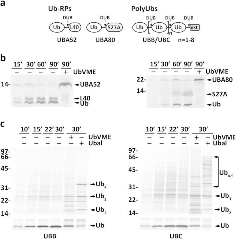
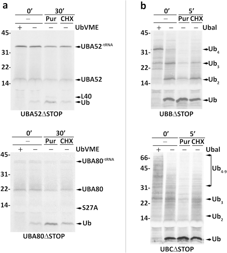
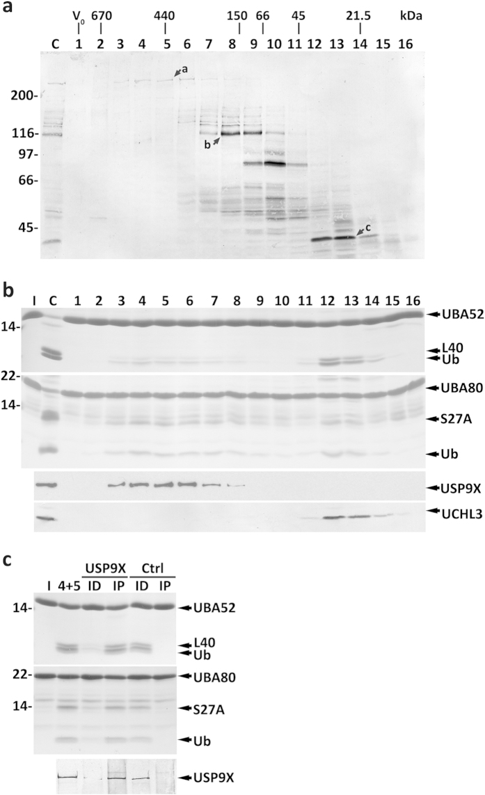
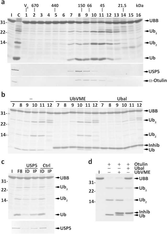
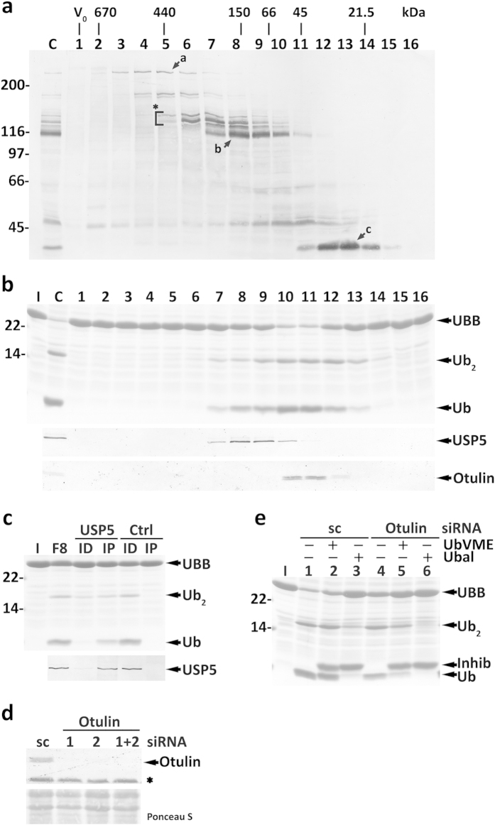
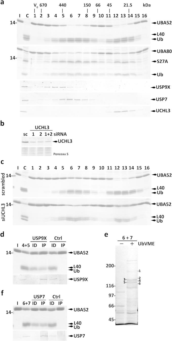
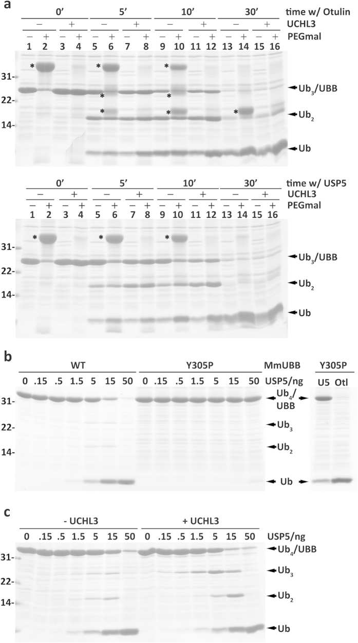
Protein ubiquitination, a major post-translational modification in eukaryotes, requires an adequate pool of free ubiquitin. Cells maintain this pool by two pathways, both involving deubiquitinases (DUBs): recycling of ubiquitin from ubiquitin conjugates and processing of ubiquitin precursors synthesized de novo. Although many advances have been made in recent years regarding ubiquitin recycling, our knowledge on ubiquitin precursor processing is still limited, and questions such as when are these precursors processed and which DUBs are involved remain largely unanswered. Here we provide data suggesting that two of the four mammalian ubiquitin precursors, UBA52 and UBA80, are processed mostly post-translationally whereas the other two, UBB and UBC, probably undergo a combination of co- and post-translational processing. Using an unbiased biochemical approach we found that UCHL3, USP9X, USP7, USP5 and Otulin/Gumby/FAM105b are by far the most active DUBs acting on these precursors. The identification of these DUBs together with their properties suggests that each ubiquitin precursor can be processed in at least two different manners, explaining the robustness of the ubiquitin de novo synthesis pathway.


Figures

Figure 1
Figure 1. Synthesis and processing of ubiquitin precursors.
(a) Schematic representation of the four mammalian ubiquitin precursors. UBA52 and UBA80 are ubiquitin-ribosomal protein fusions (Ub-RPs) in which a single ubiquitin molecule is linked to a ribosomal protein, L40 and S27A, respectively. UBB and UBC are ubiquitin polymers (polyUbs) of different lengths followed by a C-terminal extension (ext) that ranges from a single amino acid (e.g., a cysteine and a tyrosine in human and mouse UBB, respectively) to a small polypeptide (e.g., 50 amino acid residues in mouse UBC). The number of ubiquitins in mouse UBB and UBC is 4 and 9; human UBB and UBC have 3 and 9 ubiquitins, respectively. Generation of free ubiquitin from these precursors requires the action of deubiquitinases (DUB; dashed arrows). (b) 35S-labeled mouse Ub-RPs UBA52 and UBA80 and (c) mouse polyUbs UBB and UBC were synthesized in vitro in the absence or presence of DUB inhibitors (HA-UbVME or HA-Ubal), as indicated. Samples were withdrawn at the indicated time points. UBA52, UBA80, its corresponding processed products (L40, S27A and Ub), and partially processed UBB and UBC species are indicated. Note that S27A co-migrates with the abundant hemoglobin from the reticulocyte lysate and therefore is barely visible at earlier time points. In (b,c) numbers to the left indicate the molecular weights of protein standards in kDa.
Figure 2
Figure 2. Accessibility of ribosome-stalled ubiquitin precursors to DUBs.
Mouse ubiquitin precursors were in vitro synthesized using stopless mRNAs (ΔSTOP). Synthesis was for (a) 30 min or (b) 45 min, in the absence or presence of HA-UbVME or HA-Ubal, as indicated. Aliquots of synthesis reactions performed in the absence of inhibitor were further incubated in the presence of either puromycin (Pur) or cycloheximide (CHX) for the indicated time periods. In (a) the peptidyl-tRNA species (UBA52-tRNA and UBA80-tRNA), unprocessed UBA52 and UBA80, as well as the corresponding processing products, L40, S27A and ubiquitin (Ub), are indicated. Note that S27A co-migrates with the abundant hemoglobin from the reticulocyte lysate hampering its detection. In (b) partially processed UBB and UBC species are indicated. Numbers to the left indicate the molecular weights of protein standards in kDa.
Figure 3
Figure 3. Characterization of mouse liver cytosolic DUBs acting on Ub-RPs.
(a) SEC profile of HA-UbVME-reactive DUBs from mouse liver cytosol. Total cytosolic proteins (lane C) and the corresponding SEC fractions (lanes 1–16) were reacted with HA-UbVME and analyzed by SDS-PAGE/western blotting with an anti-HA antibody. Arrows a–c indicate HA-UbVME-reactive DUBs that co-elute with enzyme activities described in the main text. The elution positions of molecular mass protein standards, as well as the void volume (V0), are indicated. (b) SDS-PAGE/Coomassie blue staining analyses of UBA52 and UBA80 processing by SEC fractions (first and second panels, respectively). Lane C, total cytosolic proteins were also assayed. Ubiquitin (Ub) and the ribosomal proteins L40 and S27A are indicated. The elution profiles of USP9X and UCHL3 are also shown (lower panels). (c) Pooled fractions 4 and 5 from SEC were subjected to an immunoprecipitation/immunodepletion assay using control (lanes Ctrl) or anti-USP9X IgGs (lanes USP9X). Pooled fractions (4+5) and the corresponding immunoprecipitated (lanes IP) and immunodepleted fractions (lanes ID) were assayed for UBA52 and UBA80 cleavage (upper and middle panels, respectively). The distribution of USP9X in the samples is also shown (lower panel). Lanes I, recombinant Ub-RP fusion proteins used in the assays. Numbers to the left indicate the molecular weights of protein standards in kDa.
Figure 4
Figure 4. Characterization of mouse liver cytosolic DUBs acting on polyUbs.
(a) SDS-PAGE/Coomassie blue staining analysis of UBB processing by SEC fractions (upper panel). Lane C, total cytosolic proteins were also assayed. The elution profiles of USP5 (middle panel) and of the protein recognized by the anti-Otulin antibody (lower panel) are also shown. The elution positions of molecular mass protein standards, as well as the void volume (V0), are indicated. (b) The UBB cleavage activity comprises an HA-UbVME-sensitive and an HA-UbVME-insensitive component. SEC fractions 7–12 were preincubated in the absence or presence of HA-UbVME or HA-Ubal and used in UBB processing activity assays. Inhib, HA-UbVME or HA-Ubal. (c) SEC fraction 8 was subjected to an immunoprecipitation/immunodepletion assay using control (lanes Ctrl) or anti-USP5 IgGs (lanes USP5). Fraction 8 (F8) and the corresponding immunoprecipitated (lanes IP) and immunodepleted fractions (lanes ID) were assayed for processing activity (upper panel). The distribution of USP5 in the samples is shown (lower panel). (d) Recombinant Otulin (10 ng) pretreated or not with HA-UbVME or HA-Ubal, as indicated, was incubated with UBB for 10 min at 37 °C. A Coomassie blue-stained gel is shown. Cleavage intermediates (Ub3 and Ub2) and ubiquitin (Ub) are indicated. Lane I, recombinant mouse UBB used in the assays. Numbers to the left indicate the molecular weights of protein standards in kDa.
Figure 5
Figure 5. Characterization of HeLa cells cytosolic DUBs acting on human UBB.
(a) Total cytosolic proteins (lane C) and the corresponding SEC fractions (lanes 1–16) were reacted with HA-UbVME and analyzed by SDS-PAGE/western blot with an anti-HA antibody. The asterisk and arrows a-c indicate HA-UbVME-reactive DUBs that co-elute with enzyme activities described in the main text. The elution positions of molecular mass protein standards, as well as the void volume (V0), are indicated. (b) SDS-PAGE/Coomassie blue staining analysis of UBB processing by SEC fractions (upper panel). Lane C, total cytosolic proteins were also assayed. The elution profiles of USP5 (middle panel) and Otulin (lower panel) are also shown. (c) Fraction 8 from SEC was subjected to an immunoprecipitation/immunodepletion assay using control (lanes Ctrl) or anti-USP5 IgGs (lanes USP5). Fraction 8 (F8) and the corresponding immunoprecipitated (lanes IP) and immunodepleted fractions (lanes ID) were assayed for UBB processing activity (upper panel). The distribution of USP5 in the samples is shown (lower panel). (d) Otulin knockdown in HeLa cells. Western blot analysis of Otulin in HeLa cells transfected with Otulin-specific siRNA oligos #1 and/or #2, or with a scrambled (sc) control. The asterisk indicates a protein recognized by the anti-Otulin antibody in total homogenates, but not in cytosolic fractions. The corresponding Ponceau S-stained membrane is shown to assess protein loadings. (e) OTULIN knocked-down HeLa cell total extracts have decreased HA-UbVME-insensitive UBB processing activity. Inhib, HA-UbVME or HA-Ubal. In (b,c,e) the cleavage intermediate (Ub2), and ubiquitin (Ub) are indicated. Lanes I, recombinant UBB used in the assays. In (ac,e) numbers to the left indicate the molecular weights of protein standards in kDa.
Figure 6
Figure 6. Characterization of HeLa cells cytosolic DUBs acting on Ub-RPs.
(a) SDS-PAGE/Coomassie blue staining analyses of UBA52 and UBA80 processing by HeLa cells cytosolic SEC fractions (first and second panels, respectively). Lane C, total cytosolic proteins were also assayed. Ubiquitin (Ub) and the ribosomal proteins L40 and S27A are indicated. The elution profiles of USP9X, USP7 and UCHL3 are also shown. The protein standards used to calibrate the column, as well as the void volume (V0), are indicated. (b) Western blot analysis of UCHL3 in HeLa cells transfected with UCHL3-specific siRNA oligos #1 and/or #2, or with a scrambled (sc) control. The corresponding Ponceau S-stained membrane is shown to assess protein loadings. (c) SEC profiles of UBA52-cleavage activity in control (scrambled) and UCHL3 knocked-down (siUCHL3) HeLa cells cytosol. (d) Pooled SEC fractions 4 and 5 were subjected to an immunoprecipitation/immunodepletion assay using control (lanes Ctrl) or anti-USP9X IgGs (lanes USP9X). Pooled fractions (4 + 5) and the corresponding immunoprecipitated (lanes IP) and immunodepleted fractions (lanes ID) were assayed for UBA52 cleavage (upper panel). The distribution of USP9X in the samples is shown (lower panel). (e) Pooled fractions 6 and 7 (6 + 7) from SEC were incubated with HA-UbVME (lane +) or with HA-Ub (lane -) and subjected to immunoprecipitation using anti-HA-conjugated beads. A Coomassie blue-stained gel is shown. Numbers indicate protein bands of interest that were subjected to mass spectrometry (see Supplementary Table S1). (f) Immunoprecipitation/immunodepletion assay exactly as described in (d) but using pooled SEC fractions 6 and 7, and anti-USP7 IgGs. Lanes I in (a,c,d,f), recombinant UBA52 or UBA80, as indicated. In (a,ce) numbers to the left indicate the molecular weights of protein standards in kDa.
Figure 7
Figure 7. USP5 removes the C-terminal extension of UBB.
(a) Human UBB pretreated or not with UCHL3 was incubated with either Otulin (8 ng/lane; upper panel) or USP5 (40 ng/lane; lower panel) for the indicated periods of time. Samples were then subjected to pegylation (+PEGmal), as indicated. Pegylated species (UBB and cleavage products) are marked with asterisks. Note that in the Otulin assay, Ub2 and Ub also indicate species containing the C-terminal cysteine. (b) Mouse UBB (WT) and UBBY305P (Y305P) were incubated with increasing amounts of USP5 for 30 min at 37 °C (left panel). Processing of UBBY305P with 1 μg of USP5 (lane U5) or 10 ng of Otulin (lane Otl) is also shown (right panel). (c) Mouse UBB pretreated or not with UCHL3, as indicated, was incubated at 37 °C for 30 min with increasing amounts of USP5, as specified. Coomassie blue-stained gels are shown. Numbers to the left indicate the molecular weights of protein standards in kDa.

References

    1. Amm I., Sommer T. & Wolf D. H. Protein quality control and elimination of protein waste: the role of the ubiquitin-proteasome system. Biochim. Biophys. Acta 1843, 182–196 (2014). - PubMed
    1. van Cuijk L., Vermeulen W. & Marteijn J. A. Ubiquitin at work: The ubiquitous regulation of the damage recognition step of NER. Exp. Cell Res. 329, 101–109(2014). - PubMed
    1. Chen J. & Chen Z. J. Regulation of NF-kappaB by ubiquitination. Curr. Opin. Immunol. 25, 4–12 (2013). - PMC - PubMed
    1. Francisco T. et al. Ubiquitin in the peroxisomal protein import pathway. Biochimie 98, 29–35 (2014). - PubMed
    1. Tanno H. & Komada M. The ubiquitin code and its decoding machinery in the endocytic pathway. J. Biochem. 153, 497–504 (2013). - PubMed

Publication types

MeSH terms

LinkOut - more resources