. 2015 Aug 6;97(2):291-301.
doi: 10.1016/j.ajhg.2015.07.001. Epub 2015 Jul 30.

Mutations in TBX18 Cause Dominant Urinary Tract Malformations via Transcriptional Dysregulation of Ureter Development

Affiliations

Mutations in TBX18 Cause Dominant Urinary Tract Malformations via Transcriptional Dysregulation of Ureter Development

Asaf Vivante et al. Am J Hum Genet. .

Abstract

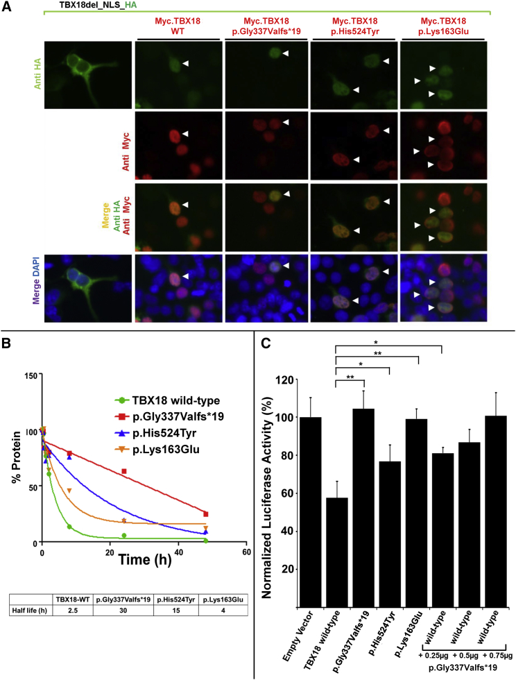
Congenital anomalies of the kidneys and urinary tract (CAKUT) are the most common cause of chronic kidney disease in the first three decades of life. Identification of single-gene mutations that cause CAKUT permits the first insights into related disease mechanisms. However, for most cases the underlying defect remains elusive. We identified a kindred with an autosomal-dominant form of CAKUT with predominant ureteropelvic junction obstruction. By whole exome sequencing, we identified a heterozygous truncating mutation (c.1010delG) of T-Box transcription factor 18 (TBX18) in seven affected members of the large kindred. A screen of additional families with CAKUT identified three families harboring two heterozygous TBX18 mutations (c.1570C>T and c.487A>G). TBX18 is essential for developmental specification of the ureteric mesenchyme and ureteric smooth muscle cells. We found that all three TBX18 altered proteins still dimerized with the wild-type protein but had prolonged protein half life and exhibited reduced transcriptional repression activity compared to wild-type TBX18. The p.Lys163Glu substitution altered an amino acid residue critical for TBX18-DNA interaction, resulting in impaired TBX18-DNA binding. These data indicate that dominant-negative TBX18 mutations cause human CAKUT by interference with TBX18 transcriptional repression, thus implicating ureter smooth muscle cell development in the pathogenesis of human CAKUT.


Figures

Figure 1
Figure 1
Identification of TBX18 Mutations in the Index Family A3880 and Three Additional Families with CAKUT (A) Pedigree of index family A3880. Squares represent males, circles females, black symbols affected persons, and white symbols unaffected persons. All affected family members presented with obstructive uropathy. Pedigree is compatible with an autosomal-dominant mode of inheritance. Roman numerals denote generations. Individuals are numbered with Roman numerals if DNA was available for study. The arrow points to the proband IV3. WT denotes the wild-type allele. p.Gly337Valfs19 indicates the mutation c.1010 deletion of G in TBX18, leading to a frameshift mutation and introducing a premature stop codon 19 codons downstream. The mutation fully segregated heterozygously (WT/p.Gly337Valfs19) across all seven affected individuals examined and was absent from all seven unaffected family members available for study (WT/WT). Red circles indicate the persons selected for whole exome sequencing. (B) Abdominal CT scan of the index individual A3880-IV3 revealing severe bilateral hydronephrosis (white asterisks) secondary to bilateral ureteral obstruction. The index individual presented during infancy and was diagnosed after evaluation of urinary tract infection. She underwent corrective surgery with multiple consecutive reconstructive operations to her lower urinary tract. She had no extrarenal manifestations. (C) Prenatal renal US of individual A3880-IV1 with bilateral hydronephrosis at a gestational age of 28 weeks. Postnatally, he had unilateral pelviectasis (Figure S1A) with normal voiding cysturethrogram. At the age of 6.5 years there was left-sided renal hypodysplasia with no signs of obstruction (Figures S1B and S1C). (D) Exon structure of human TBX18 cDNA and domain structure of the T-box domain 18 (TBX18) protein. TBX18 contains a nuclear localization signal (NLS, red); an engrailed homology-1 motif (EH1, blue), and a DNA-binding T-box domain (yellow). Start codon (ATG) and stop codon (TGA) are indicated. (E) Chromatograms of heterozygous mutations detected in TBX18 (in relation to exons and protein domains) in the index family (red) and three additional families (black) with CAKUT. The index family’s heterozygous mutation c.1010delG leads to a frameshift and after 19 amino acids to a premature termination codon (p.Gly337Valfs19).
Figure 2
Figure 2
Trafficking of TBX18 to Nuclei via Its Nuclear Localization Signal, Prolonged Half-Life of Mutant Proteins, and Lack of Transcriptional Repression by TBX18 Mutants (A) Immunofluorescence images in HEK293 cells that were cotransfected with an HA-tagged construct (TBX18del_NLS_HA) that lacks the nuclear localization signal (green), together with either (red) a human Myc-tagged labeled TBX18 wild-type construct (Myc.TBX18_WT) or constructs representing the substitution of family A3880 (Myc.TBX18_Gly337Valfs19), the substitution of families A2900 and A2385 (Myc.TBX18_His524Tyr), or the substitution of family ICH_006 (Myc.TBX18_Lys163Glu). Note that the NLS-deficient construct TBX18del_NLS_HA fails to localize to the nucleus (left column). However, in the presence of co-transfection of either Myc.TBX18_WT or either altered protein (red), even the NLS-deficient proteins (green) traffic to the nucleus, most likely by T-box-mediated dimerization with the NLS-containing Myc-tagged construct (red). Double-transfected cells (HA-construct and Myc-construct) are denoted by arrowheads. (B) Half-life estimation of wild-type and mutant TBX18. Although the half-life of wild-type TBX18 was determined to be 2.5 hr in vitro, the half-lives of the mutant proteins were prolonged to more than 30 hr (p.Gly337Valfs19), 15 hr (p.His524Tyr), and 4 hr (p.Lys163Glu). (C) Luciferase transcriptional reporter assay for TBX18 wild-type and mutant constructs. Compared to empty vector control (set at 100% of luciferase activity), transfection with TBX18 wild-type construct causes transcriptional repression to 58%. All three mutant constructs (p.Gly337Valfs19, p.His524Tyr, and p.Lys163Glu) detected in CAKUT-affected families repressed transcriptional activity significantly less than the wild-type construct. Co-transfection of TBX18 wild-type construct and the protein truncating construct p.Gly337Valfs19 in increasing amounts yielded a dose-dependent lack of repression for the mutant construct, supporting its dominant-negative effect on transcriptional repression by the wild-type construct. p < 0.05, ∗∗p < 0.01.
Figure 3
Figure 3
Three-Dimensional Model of Crystal Structure of Human TBX18 Paralog TBX1 in Complex with Cognate DNA (A) Ribbon diagram of the TBX1-DNA complex in orthogonal views. Depicted are residues of two TBX18 monomers. β-pleated sheets are marked as yellow arrows, α helices in red, and DNA is marked in white as space-filling model. Residues marked in blue are those found at the protein-DNA interface (see B). Arrows point to the position of the amino acid residue Lys163 (red) found altered (p.Lys163Glu) in family ICH_006 with CAKUT. Inset on the right shows that Lys163 (arrow pointing to red Lys163 side chain) is in direct vicinity to the DNA interaction interface (white) (modeled in TBX1, PDB: 4A04). (B) Partial sequence alignment of TBX18 T-box domain and its paralogs, for which protein 3D crystal structure data are available (H. sapiens TBX1, TBX3, and TBX5 and X. laevis brachyury). Amino acid residue numbers are shown per alignment row. Residues in blue are in the vicinity of the TBX1 protein-DNA interface (see A). Residue Lys163 found altered (p.Lys163Glu) in family ICH_006 with CAKUT is shown in red. (C) DNA binding assay of wild-type TBX18 and TBX18 mutants detected in individuals with CAKUT show that p.Lys163Glu substitution fails to bind to DNA. Electrophoretic mobility shift assay (EMSA) was performed with equal amounts of in vitro synthesized myc-tagged constructs Myc.TBX18_wild-type (wild-type), Myc.TBX18_Gly337Valfs19, Myc.TBX18_His524Tyr, or Myc.TBX18_Lys163Glu. All alleles except for p.Lys163Glu bind to the TBX18 DNA target site as indicated by retardation of the labeled probe (red arrowheads in lanes 3). Specificity of the binding is confirmed by the addition of anti-myc antibody that results in formation of a super shifted complex (black arrowheads in lanes 5).

References

    1. Smith J.M., Stablein D.M., Munoz R., Hebert D., McDonald R.A. Contributions of the Transplant Registry: The 2006 Annual Report of the North American Pediatric Renal Trials and Collaborative Studies (NAPRTCS) Pediatr. Transplant. 2007;11:366–373. - PubMed
    1. Chen F. Genetic and developmental basis for urinary tract obstruction. Pediatr. Nephrol. 2009;24:1621–1632. - PMC - PubMed
    1. Woolf A.S. A molecular and genetic view of human renal and urinary tract malformations. Kidney Int. 2000;58:500–512. - PubMed
    1. Pohl M., Bhatnagar V., Mendoza S.A., Nigam S.K. Toward an etiological classification of developmental disorders of the kidney and upper urinary tract. Kidney Int. 2002;61:10–19. - PubMed
    1. Sanna-Cherchi S., Sampogna R.V., Papeta N., Burgess K.E., Nees S.N., Perry B.J., Choi M., Bodria M., Liu Y., Weng P.L. Mutations in DSTYK and dominant urinary tract malformations. N. Engl. J. Med. 2013;369:621–629. - PMC - PubMed

Publication types