. 2015 Aug 20;59(4):677-84.
doi: 10.1016/j.molcel.2015.06.029. Epub 2015 Jul 30.

Pin1-Induced Proline Isomerization in Cytosolic p53 Mediates BAX Activation and Apoptosis

Affiliations

Pin1-Induced Proline Isomerization in Cytosolic p53 Mediates BAX Activation and Apoptosis

Ariele Viacava Follis et al. Mol Cell. .

Abstract

The cytosolic fraction of the tumor suppressor p53 activates the apoptotic effector protein BAX to trigger apoptosis. Here we report that p53 activates BAX through a mechanism different from that associated with activation by BH3 only proteins (BIM and BID). We observed that cis-trans isomerization of proline 47 (Pro47) within p53, an inherently rare molecular event, was required for BAX activation. The prolyl isomerase Pin1 enhanced p53-dependent BAX activation by catalyzing cis-trans interconversion of p53 Pro47. Our results reveal a signaling mechanism whereby proline cis-trans isomerization in one protein triggers conformational and functional changes in a downstream signaling partner. Activation of BAX through the concerted action of cytosolic p53 and Pin1 may integrate cell stress signals to induce a direct apoptotic response.


Figures

Figure 1
Figure 1. BAX activation by p53 is mediated by the cis isomer of p53 Pro47 and enhanced by the prolyl isomerase Pin1 (see also Figure S1)
A. p53 constructs. From top to bottom: full-length p53, with domain boundaries; near full-length recombinant construct (residues 1–360, purple edges); transactivation domain (NTD, residues 1–102, red); DNA binding domain (DBD, residues 102–312, blue); combination of NTD and DBD (N–D, residues 1–292, cyan edges); combination of DBD and tetramerization domain (D–T, residues 102–360, brown edges). B. BAX-dependent permeabilization of large unlilamellar vesicles (LUVs), after one hour incubation with p53 constructs or the bona fide BAX activator, n/c-BID. Full-length p53 and shorter p53 constructs containing the NTD segment activated BAX. Mean values +/− SEM; N = 5. C. Perturbation of the 1H-15N HSQC spectrum of p53-NTD (red) upon titration of unlabeled BAX (olive, left panel) or GST-Pin1 (orange to olive gradient, right panel). Titration of BAX resulted in the appearance of slow exchanging satellite peaks for resonances of residues surrounding p53 Pro47. Titration of Pin1 resulted in a similar chemical shift perturbation (CSP) pattern that transitioned to a fast-exchange regime at increasing Pin1 concentrations. The insets at the top right of each panel show an equivalent perturbation effect in both titrations for the Cα-CO resonance of p53 Pro47. D. Oligomerization of BAX in LUV samples, detected by chemical crosslinking, in the presence of p531-360 (5–100 nM) or NTD and in the absence or presence of Pin1. E. LUV permeabilization induced by 100 nM BAX and p531-360 (purple) or p53-NTD (red), at increasing Pin1 concentrations (110 nM – 1.5 μM; left), and (right) in a separate assay, in the presence of p531-360 and wild type or inactive Pin1 C113S mutant (both 1 μM). Mean +/− SEM; *P<0.05; **P<0.01.
Figure 2
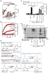
Figure 2. Insights into the mechanism of p53-mediated BAX activation: enhancement by an optimized Pin1 recognition motif in the NTD of p53 and requirement for the ability of p53 Pro47 to undergo isomerization (See also Figures S1, S2)
A. Perturbation of the 1H-15N HSQC spectrum of phosphomimetic p53-NTD S46E mutant (burgundy) upon titration of unlabeled Pin1 (orange to olive gradient). Pin1 induced CSPs in resonances of residues adjacent to Glu46-Pro47. B. Kinetic measurements of BAX-dependent LUV permeabilization in the presence of wild type p53-NTD (red) or p53-NTD S46E (burgundy, both 200 nM) in the absence (crosses) and presence (circles) of 1μM Pin1. C. BAX-dependent LUV permeabilization in the presence of synthetic peptides encompassing p53 residues 40 – 59 and containing phosphorylated Ser46, a P47S mutation, or non natural Pro47 derivatives with altered cis-trans isomerization ratios and/or rates (captions are color coded to match the schemes in panel E), in the absence (white bars) and presence of Pin1 (black bars). Mean +/− SEM; **P<0.01; ***P<0.001. D. BAX oligomerization in LUV samples treated with p5340-59 peptides and Pin1 probed by chemical crosslinking. E. Structures and cis-trans isomerization properties of the non-natural proline derivatives incorporated in p5340-59 peptides. Compared to proline, the cis isomer is dominant in 5,5 dimethyl-proline (5,5-DMP); 4,4-difluoro-proline (4,4-F2P) has faster isomerization rate; (4S)-fluoro-proline [(4S)-FP] exhibits slightly accelerated isomerization and higher cis content, while in (4R)-fluoro-proline [(4R)-FP] the trans isomer is stabilized. The indicated ratios and rates were reported in (An et al., 1999) or (Renner et al., 2001). In p5340-59 peptides we determined the content of trans and cis Pro47 or 5,5-DMP47 from the intensity of corresponding β and γ side-chain resonances in 1H13C-HSQC spectra.
Figure 3
Figure 3. Mapping of binding interfaces between BAX and p53 (see also Figures S2, S3)
A. Perturbation of the 1H-15N HSQC spectrum of BAX (grey) upon titration of unlabeled p531-360 (purple). Select resonances that exhibited noticeable CSP or line broadening are labeled. B. Structure of BAX showing residues that exhibited CSP upon titration of unlabeled p531-360, color-coded according to the observed perturbation of individual residues upon titration of isolated p53-NDT (red), p53-DBD (blue) or both domains (purple). The positions of residues indicated in panel A are highlighted. C. Perturbation of the 1H-15N HSQC spectrum of p53-DBD (blue) upon titration of unlabeled BAX (olive). This domain of p53 interacted with BAX without directly promoting its activation. D. Structure mapping of p53-DBD residues whose resonances experienced significant CSP upon titration of unlabeled BAX (olive spheres). The positions of residues indicated in panel C are highlighted. E. A proposed model for the simultaneous interaction of p53-NTD (residues 40–59) and p53-DBD with BAX. The disordered segment connecting the two interacting regions is shown as a dashed line. The Ser46-Pro47 motif within p53-NTD is indicated. F. Schematic representation of interactions between BAX (grey) and BH3 activators (purple, orange), and the interaction of BAX with p53-NTD (red) and p53-DBD (blue) identified in this study. The purple helical segment, representing a ‘stapled’ BIM BH3 peptide, is bound to a α1-α6 ‘allosteric’ site on BAX (Gavathiotis et al., 2008). Upon engagement of this site, BAX α9 (transparent) is displaced to enable binding of BH3 activators to the ‘canonical’ α3-5 site (the orange helical segment represents BID BH3 bound to this site) (Czabotar et al., 2013) and thus trigger complete BAX activation through the further displacement of α6-α8 from the protein core. Based on the position of the p53-NTD (red) interaction site on BAX, ‘bridging’ the two BH3 binding sites, it appears likely that cis-trans isomerization of p53 Pro47 could trigger the simultaneous displacement of BAX α6-α9 in a single step as opposed to the ‘two step’ activation mechanism postulated for BAX activation by BH3 proteins.
Figure 4
Figure 4. Pin1-mediated activation of BAX by cytosolic p53 in cells and proposed mechanism of activation (see also Figure S4)
A. Western blots showing expression levels of endogenous or stably over-expressed Pin1 and tamoxifen-inducible, estrogen receptor fusion wild type p53 (p53-ERTam) and mutations of its Pin1-consensus – BAX activation site in p53-null background H1299 cells. B. Co-immunoprecipitation between p53-ERTam (wild type, mutant) and Pin1. C. Fluorescence activated cell sorting (FACS) analysis of H1299 p53-ERTam cells with endogenous (grey bars) or over-expressed Pin1 (black bars) after UV irradiation. Only Pin1 over-expressing cells showed sustained apoptosis, measured by Annexin V staining, upon 4-hydroxy-tamoxifen (4-OHT) activation of wild type p53-ERTam in the presence of the protein synthesis inhibitor cycloheximide (CHX). Mean +/− SEM; *P<0.05; **P<0.01. D. Annexin V staining and FACS analysis of H1299 p53-ERTam wild type cells with endogenous (grey bars) or siRNA-silenced Pin1 (light blue bars) after UV irradiation. Pin1 RNAi reduced apoptosis after p53-ERTam activation both in the absence and presence of CHX. Pin1 clearance upon RNAi was confirmed by western blot (inset). Mean +/− SEM; **P<0.01. E. Annexin V staining and FACS analysis of H1299 p53-ERTam wild type cells over-expressing Pin1 after siRNA silencing of BAK (grey bars) or BAX (orange bars) expression followed by UV irradiation. BAX but not BAK knockdown reduced the levels of Annexin V staining, indicating that apoptosis in these cells depends mainly on BAX activation. Western blots detecting BAK and BAX levels after RNAi are shown on the right. Mean +/− SEM; ***P<0.001. F. Annexin V staining and FACS analysis of p53 null H1299 cells after siRNA-silencing of BAX (in Pin1 over-expressing cells; left side; orange bars) or Pin1 (in endogenous Pin1 cells; right side, light blue bars) followed by transfection of p5340-59 peptides. Mean +/− SEM; ***P<0.001. G. Cartoon of the proposed mechanism by which p53 activates BAX in concert with Pin1. Structure of BAX with the segments involved in activation by BH3 proteins highlighted in purple and orange (left). Illustration of the Ser46-Pro47 motif and flanking regions of p53-NTD, upon cis isomerization of Pro47, interacting with BAX (center). This event may be favored by prior Pin1-catalyzed isomerization of p53 Pro47. Pin1 catalyzed or spontaneous cis-trans isomerization of BAX-bound p53 Pro47 is proposed to trigger conformational rearrangements in the neighboring residues of p53 and consequently destabilize the C-terminal α-helices of BAX that contact this p53 segment, leading to BAX activation (right).

Similar articles

Cited by

References

    1. An SSA, Lester CC, Peng JL, Li YJ, Rothwarf DM, Welker E, Thannhauser TW, Zhang LS, Tam JP, Scheraga HA. Retention of the cis proline conformation in tripeptide fragments of bovine pancreatic ribonuclease A containing a non-natural proline analogue, 5,5-dimethylproline. J Am Chem Soc. 1999;121:11558–11566.
    1. Asciolla JJ, Renault TT, Chipuk JE. Examining BCL-2 family function with large unilamellar vesicles. J Vis Exp. 2012;68:4291. - PMC - PubMed
    1. Berjanskii MV, Neal S, Wishart DS. PREDITOR: a web server for predicting protein torsion angle restraints. Nucleic Acids Res. 2006;34:W63–W69. - PMC - PubMed
    1. Chipuk JE, Bouchier-Hayes L, Kuwana T, Newmeyer DD, Green DR. PUMA couples the nuclear and cytoplasmic proapoptotic function of p53. Science. 2005;309:1732–1735. - PubMed
    1. Chipuk JE, Kuwana T, Bouchier-Hayes L, Droin NM, Newmeyer DD, Schuler M, Green DR. Direct activation of Bax by p53 mediates mitochondrial membrane permeabilization and apoptosis. Science. 2004;303:1010–1014. - PubMed

Publication types

MeSH terms

LinkOut - more resources