. 2015 Jul 14:9:102.
doi: 10.3389/fnsys.2015.00102. eCollection 2015.

Age-related changes in dentate gyrus cell numbers, neurogenesis, and associations with cognitive impairments in the rhesus monkey

Affiliations

Age-related changes in dentate gyrus cell numbers, neurogenesis, and associations with cognitive impairments in the rhesus monkey

Laura B Ngwenya et al. Front Syst Neurosci. .

Abstract

The generation of new neurons in the adult mammalian brain is well-established for the hippocampal dentate gyrus (DG). However, the role of neurogenesis in hippocampal function and cognition, how it changes in aging, and the mechanisms underlying this are yet to be elucidated in the monkey brain. To address this, we investigated adult neurogenesis in the DG of 42 rhesus monkeys (39 cognitively tested) ranging in age from young adult to the elderly. We report here that there is an age-related decline in proliferation and a delayed development of adult neuronal phenotype. Additionally, we show that many of the new neurons survive throughout the lifetime of the animal and may contribute to a modest increase in total neuron number in the granule cell layer of the DG over the adult life span. Lastly, we find that measures of decreased adult neurogenesis are only modestly predictive of age-related cognitive impairment.

Keywords: adult neurogenesis; bromodeoxyuridine; cognitive aging; dentate gyrus; doublecortin; learning; non-human primate; stereology.


Figures

FIGURE 1
FIGURE 1
Capacity for neurogenesis declines with age. (A) The total number of Ki-67 positive nuclei significantly declines with age. Regression analysis predicts a 68% decline in Ki-67 positive cells between a 7 and a 25-years-old monkey (threefold change). (B) The total number of BrdU positive cell nuclei that are present after a 3-weeks survival also shows a significant negative correlation with age. Regression predicts a 53% decline between ages 7 and 25, which corresponds to a twofold change in BrdU labeled cells. (C) A photomicrograph illustrates BrdU immunohistochemistry with cresyl violet counterstain in the DG of a young monkey; scale bar = 100μm. The box represents a cluster of BrdU positive nuclei, which is enlarged in (D). (E) Aged animals also show clusters of BrdU positive nuclei as shown here. Scale bar for (D,E) = 20 μm.
FIGURE 2
FIGURE 2
Total number of DCX positive cells in the DG declines sharply with age. More DCX positive cells are seen in the granule cell layer of the DG in young animals than in old animals. (A) DCX positive cells in a 7.9 years-old animal. (B) DCX positive cells in a 24.5 years-old animal. Scale bar for (A,B) = 20 μm. (C) There is a significant decline in the number of DCX positive cells present with increasing age.
FIGURE 3
FIGURE 3
Newly generated cells differentiate into mature neurons, however, the process may be delayed in aged animals. (A) A BrdU (green) and DCX (red) double labeled cell in the hilus of the DG of a 6.9 years monkey at 3 weeks post-BrdU injection. (B) Most BrdU and DCX positive cells are seen in the GCL, as in this 8 years-old monkey with a 38-weeks survival time. (C) New mature neurons, as labeled with both BrdU (green) and NeuN (red) are seen in the GCL of animals with survival times longer than 1 year, as in this 9.2 years-old animal with an 82-weeks survival time. (D) Old animals also continue to have survival of new neurons as in this 19.9 yr old monkey with an 83-weeks post-BrdU survival time. Scale bars in (A–D) = 20 μm. (E) Immature neurons are sparse in the hilus, especially at longer survival times. (F) The majority of BrdU and DCX double labeled cells are found in the GCL. Whereas young animals average 25% BrdU positive cells express DCX, no immature neurons are seen in old animals at 3 weeks. With prolonged survival time in young animals, the percentage of BrdU positive DCX double labeled cells declines, yet in aged animals, BrdU and DCX double labeled cells are just beginning to be seen with longer survival times. (G) As survival times post-BrdU approach 1 year and beyond, both young and old animals show a fairly steady percentage of new mature neurons as seen by percentage of BrdU labeled and NeuN double labeled cells in the GCL. (H) In animals with post-BrdU survival times between 43 and 83 weeks, the percentage of BrdU and NeuN double labeled cells declines with age.
FIGURE 4
FIGURE 4
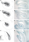
Regions of interest for stereological evaluation of hippocampal cell numbers and volumes. The entire rostral-caudal extent of the hippocampus was evaluated. Representative schematics (A,C,E,G) and photomicrographs (B,D,F,H) from rostral to caudal thionin stained sections are shown. Scale bar = 1 mm. The granule cell layer (GCL), molecular layer (ML), and hilus of the DG were analyzed separately and defined as outlined. For consistency of ROI, the hilus was defined as that area in between the blades of the granule cell layer. The “remainder of the hippocampus” included that area which represents CA3, CA2, and CA1, as outlined.
FIGURE 5
FIGURE 5
Stereological analysis of DG shows increases with age. (A) The estimated total number of granule cell neurons in the hippocampal DG increases over the lifespan of the rhesus monkey. This corresponds to a volume increase in the DG (B). However, the remainder of the hippocampus, which includes non-neurogenic CA3, CA2, and CA1 does not show a corresponding increase with age (C).
FIGURE 6
FIGURE 6
Cognitive decline in aging animals. (A) In the hippocampal dependent task DNMS with 10-min delay, the percentage of correct responses decreased with increasing age (r = -0.503, p = 0.002). A similar response was seen in the DRST task, both spatial and object conditions. The total span obtained in DRST object (B) and DRST spatial (C) was negatively correlated with age (p = 0.027, p = 0.001; respectively). (D) In CSST, the number of trials required to master initial abstraction increased with age (r = 0.365, p = 0.040). (E) The total number of broken sets (r = 0.413, p = 0.019) and (F) the total number of perseverative errors also showed a positive correlation with increasing age (r = 0.349, p = 0.050).
FIGURE 7
FIGURE 7
Short-term survival of newly generated cells shows correlation with the cognitive task CSST. (A) Schematic of the CSST task in which each box represents a trial; the asterisk represents the correct answer. Animals first learn that “red” is correct, as shown in the first row of trials. After ten consecutive correct responses, the correct response shifts to the new concept of “triangle”, and a new set of trials begins, as illustrated. (B) The total number of BrdU positive cells in the DG after a 3 weeks survival was negatively correlated with the number of trials needed for initial abstraction (r = -0.737, p = 0.015). (C) The total number of broken sets was also negatively correlated with BrdU number (r = -0.741, p = 0.014). (D) The total number of perseverative errors showed a trend, but was not significantly correlated with the total number of BrdU cells present in the DG at 3 weeks (r = -0.550, p = 0.100).

Similar articles

Cited by

References

    1. Aggleton J. P., Wright N. F., Rosene D. L., Saunders R. C. (2015). Complementary patterns of direct amygdala and hippocampal projections to the macaque prefrontal cortex. Cereb. Cortex 10.1093/cercor/bhv019 [Epub ahead of print]. - DOI - PMC - PubMed
    1. Aimone J. B., Deng W., Gage F. H. (2010). Adult neurogenesis: integrating theories and separating functions. Trends Cogn. Sci. 14 325–337. 10.1016/j.tics.2010.04.003 - DOI - PMC - PubMed
    1. Aizawa K., Ageyama N., Terao K., Hisatsune T. (2011). Primate-specific alterations in neural stem/progenitor cells in the aged hippocampus. Neurobiol. Aging 32 140–150. 10.1016/j.neurobiolaging.2008.12.011 - DOI - PubMed
    1. Aizawa K., Ageyama N., Yokoyama C., Hisatsune T. (2009). Age-dependent alteration in hippocampal neurogenesis correlates with learning performance of macaque monkeys. Exp. Anim. 58 403–407. 10.1538/expanim.58.403 - DOI - PubMed
    1. Altman J., Das G. D. (1965). Autoradiographic and histological evidence of postnatal hippocampal neurogenesis in rats. J. Comp. Neurol. 124 319–335. 10.1002/cne.901240303 - DOI - PubMed

LinkOut - more resources