

Decoding illusory self-location from activity in the human hippocampus
- PMID: 26236222
- PMCID: PMC4502353
- DOI: 10.3389/fnhum.2015.00412
Decoding illusory self-location from activity in the human hippocampus
Abstract
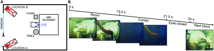
Decades of research have demonstrated a role for the hippocampus in spatial navigation and episodic and spatial memory. However, empirical evidence linking hippocampal activity to the perceptual experience of being physically located at a particular place in the environment is lacking. In this study, we used a multisensory out-of-body illusion to perceptually 'teleport' six healthy participants between two different locations in the scanner room during high-resolution functional magnetic resonance imaging (fMRI). The participants were fitted with MRI-compatible head-mounted displays that changed their first-person visual perspective to that of a pair of cameras placed in one of two corners of the scanner room. To elicit the illusion of being physically located in this position, we delivered synchronous visuo-tactile stimulation in the form of an object moving toward the cameras coupled with touches applied to the participant's chest. Asynchronous visuo-tactile stimulation did not induce the illusion and served as a control condition. We found that illusory self-location could be successfully decoded from patterns of activity in the hippocampus in all of the participants in the synchronous (P < 0.05) but not in the asynchronous condition (P > 0.05). At the group-level, the decoding accuracy was significantly higher in the synchronous than in the asynchronous condition (P = 0.012). These findings associate hippocampal activity with the perceived location of the bodily self in space, which suggests that the human hippocampus is involved not only in spatial navigation and memory but also in the construction of our sense of bodily self-location.
Keywords: body perception; multisensory integration; perceptual illusion; self-consciousness; self-location.
Figures


Similar articles
-
Duplication of the bodily self: a perceptual illusion of dual full-body ownership and dual self-location.R Soc Open Sci. 2020 Dec 9;7(12):201911. doi: 10.1098/rsos.201911. eCollection 2020 Dec. R Soc Open Sci. 2020. PMID: 33489299 Free PMC article.
-
Peripersonal space as the space of the bodily self.Cognition. 2015 Nov;144:49-57. doi: 10.1016/j.cognition.2015.07.012. Epub 2015 Jul 29. Cognition. 2015. PMID: 26231086 Free PMC article.
-
The Redundant Signals Effect and the Full Body Illusion: not Multisensory, but Unisensory Tactile Stimuli Are Affected by the Illusion.Multisens Res. 2021 Apr 9:1-33. doi: 10.1163/22134808-bja10046. Online ahead of print. Multisens Res. 2021. PMID: 33838624
-
Bodily ownership and self-location: components of bodily self-consciousness.Conscious Cogn. 2013 Dec;22(4):1239-52. doi: 10.1016/j.concog.2013.08.013. Epub 2013 Sep 13. Conscious Cogn. 2013. PMID: 24025475 Review.
-
The vestibular system: a spatial reference for bodily self-consciousness.Front Integr Neurosci. 2014 Apr 17;8:31. doi: 10.3389/fnint.2014.00031. eCollection 2014. Front Integr Neurosci. 2014. PMID: 24860446 Free PMC article. Review.
Cited by
-
A Protective Mechanism against Illusory Perceptions Is Amygdala-Dependent.J Neurosci. 2019 Apr 24;39(17):3301-3308. doi: 10.1523/JNEUROSCI.2577-18.2019. Epub 2019 Feb 25. J Neurosci. 2019. PMID: 30804094 Free PMC article.
-
Virtual Enactment Effect on Memory in Young and Aged Populations: a Systematic Review.J Clin Med. 2019 May 7;8(5):620. doi: 10.3390/jcm8050620. J Clin Med. 2019. PMID: 31067784 Free PMC article. Review.
-
Direct Electrophysiological Correlates of Body Ownership in Human Cerebral Cortex.Cereb Cortex. 2019 Mar 1;29(3):1328-1341. doi: 10.1093/cercor/bhy285. Cereb Cortex. 2019. PMID: 30496342 Free PMC article.
-
Activity of the inferior parietal cortex is modulated by visual feedback delay in the robot hand illusion.Sci Rep. 2019 Jul 11;9(1):10030. doi: 10.1038/s41598-019-46527-8. Sci Rep. 2019. PMID: 31296914 Free PMC article.
-
Neural bases of the bodily self as revealed by electrical brain stimulation: A systematic review.Hum Brain Mapp. 2023 May;44(7):2936-2959. doi: 10.1002/hbm.26253. Epub 2023 Feb 28. Hum Brain Mapp. 2023. PMID: 36852645 Free PMC article.
References
-
- Andersen P. (2007). The Hippocampus Book. Oxford: Oxford University Press.
LinkOut - more resources
Full Text Sources
Other Literature Sources