

Modifications of myofilament protein phosphorylation and function in response to cardiac arrest induced in a swine model
- PMID: 26236240
- PMCID: PMC4503891
- DOI: 10.3389/fphys.2015.00199
Modifications of myofilament protein phosphorylation and function in response to cardiac arrest induced in a swine model
Abstract
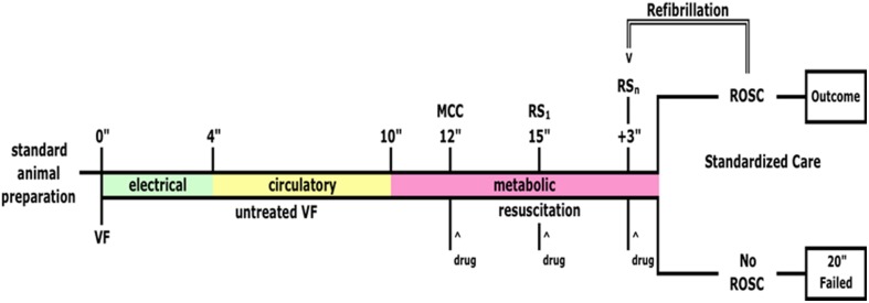
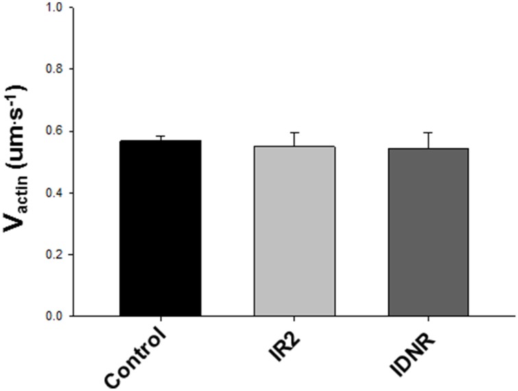
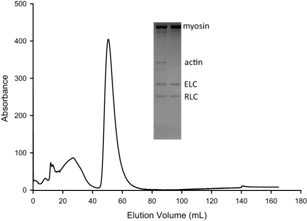
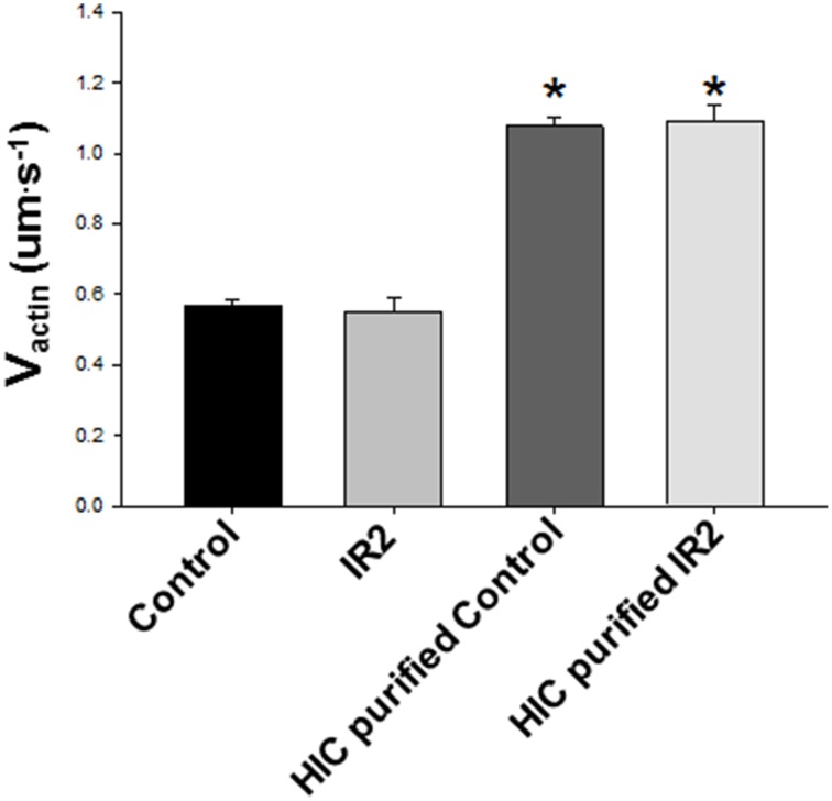
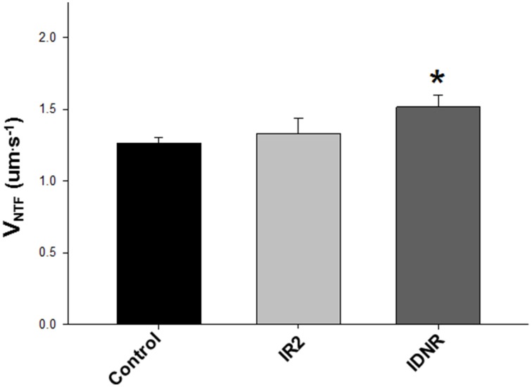
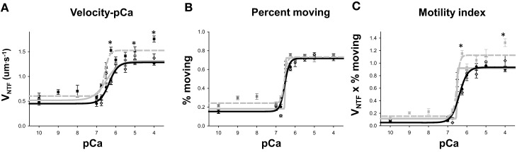
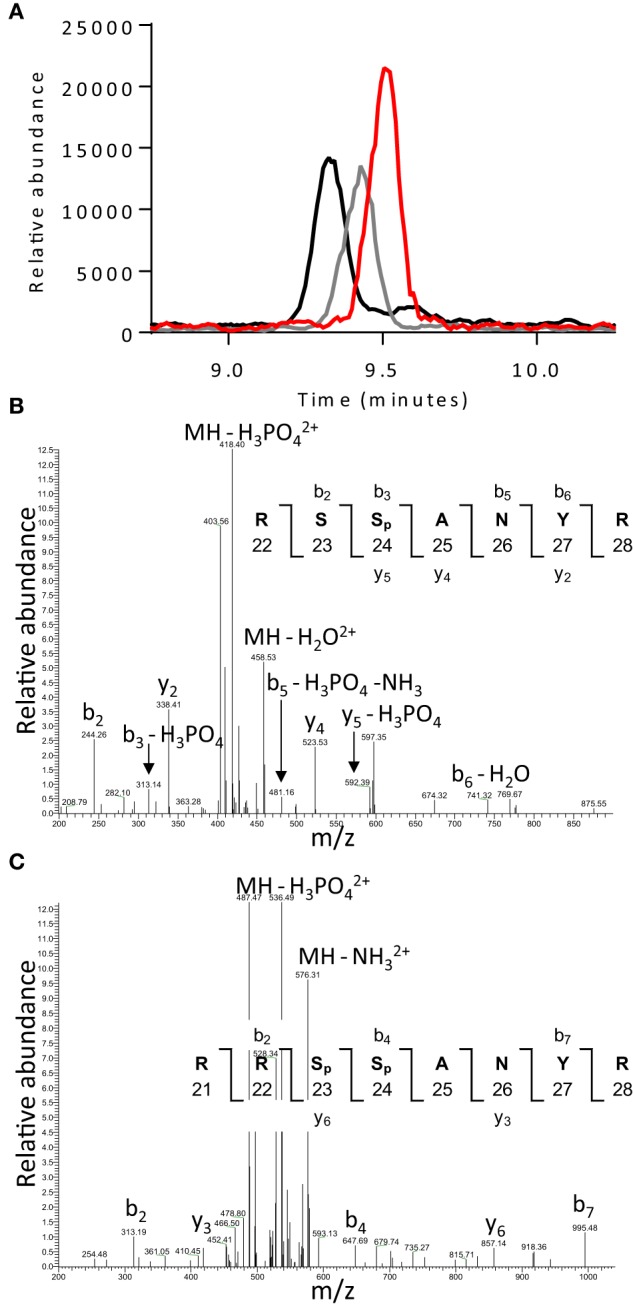
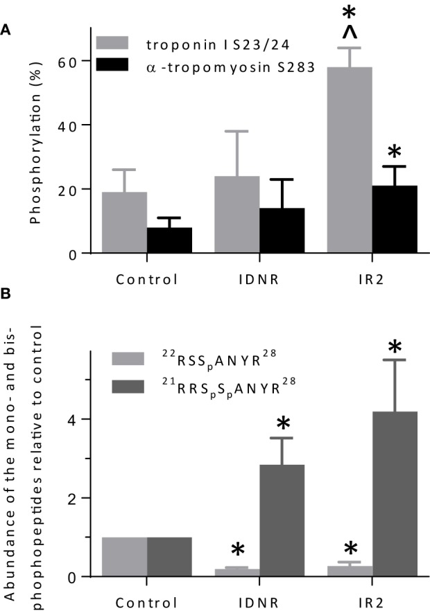
Cardiac arrest is a prevalent condition with a poor prognosis, attributable in part to persistent myocardial dysfunction following resuscitation. The molecular basis of this dysfunction remains unclear. We induced cardiac arrest in a porcine model of acute sudden death and assessed the impact of ischemia and reperfusion on the molecular function of isolated cardiac contractile proteins. Cardiac arrest was electrically induced, left untreated for 12 min, and followed by a resuscitation protocol. With successful resuscitations, the heart was reperfused for 2 h (IR2) and the muscle harvested. In failed resuscitations, tissue samples were taken following the failed efforts (IDNR). Actin filament velocity, using myosin isolated from IR2 or IDNR cardiac tissue, was nearly identical to myosin from the control tissue in a motility assay. However, both maximal velocity (25% faster than control) and calcium sensitivity (pCa50 6.57 ± 0.04 IDNR vs. 6.34 ± 0.07 control) were significantly (p < 0.05) enhanced using native thin filaments (actin+troponin+tropomyosin) from IDNR samples, suggesting that the enhanced velocity is mediated through an alteration in muscle regulatory proteins (troponin+tropomyosin). Mass spectrometry analysis showed that only samples from the IR2 had an increase in total phosphorylation levels of troponin (Tn) and tropomyosin (Tm), but both IR2 and IDNR samples demonstrated a significant shift from mono-phosphorylated to bis-phosphorylated forms of the inhibitory subunit of Tn (TnI) compared to control. This suggests that the shift to bis-phosphorylation of TnI is associated with the enhanced function in IDNR, but this effect may be attenuated when phosphorylation of Tm is increased in tandem, as observed for IR2. There are likely many other molecular changes induced following cardiac arrest, but to our knowledge, these data provide the first evidence that this form cardiac arrest can alter the in vitro function of the cardiac contractile proteins.
Keywords: cardiac arrest; motility; myosin; phosphorylation; resuscitation; troponin.
Figures
Similar articles
-
Regulation of contraction in striated muscle.Physiol Rev. 2000 Apr;80(2):853-924. doi: 10.1152/physrev.2000.80.2.853. Physiol Rev. 2000. PMID: 10747208 Review.
-
Expression of troponin subunits in the rat renal afferent arteriole.IUBMB Life. 2019 Oct;71(10):1475-1481. doi: 10.1002/iub.2061. Epub 2019 May 2. IUBMB Life. 2019. PMID: 31046198
-
Cardiac actin capping protein reduction and protein kinase C inhibition maintain myofilament function during cardioplegic arrest.Cell Physiol Biochem. 2011;27(3-4):263-72. doi: 10.1159/000327952. Epub 2011 Apr 1. Cell Physiol Biochem. 2011. PMID: 21471715
-
Investigation of the effects of phosphorylation of rabbit striated muscle alpha alpha-tropomyosin and rabbit skeletal muscle troponin-T.Eur J Biochem. 1994 Apr 1;221(1):129-37. doi: 10.1111/j.1432-1033.1994.tb18721.x. Eur J Biochem. 1994. PMID: 8168502
-
Regulation of contractile proteins in diabetic heart.Cardiovasc Res. 1997 Apr;34(1):34-40. doi: 10.1016/s0008-6363(97)00059-x. Cardiovasc Res. 1997. PMID: 9217870 Review.
Cited by
-
Early-Stage Alcoholic Cardiomyopathy Highlighted by Metabolic Remodeling, Oxidative Stress, and Cardiac Myosin Dysfunction in Male Rats.Int J Mol Sci. 2025 Jul 15;26(14):6766. doi: 10.3390/ijms26146766. Int J Mol Sci. 2025. PMID: 40725013 Free PMC article.
-
Impact of regulatory light chain mutation K104E on the ATPase and motor properties of cardiac myosin.J Gen Physiol. 2021 Jul 5;153(7):e202012811. doi: 10.1085/jgp.202012811. J Gen Physiol. 2021. PMID: 33891674 Free PMC article.
-
Modeling thick filament activation suggests a molecular basis for force depression.Biophys J. 2024 Mar 5;123(5):555-571. doi: 10.1016/j.bpj.2024.01.024. Epub 2024 Feb 1. Biophys J. 2024. PMID: 38291752 Free PMC article.
-
Acidosis modifies effects of phosphorylated tropomyosin on the actin-myosin interaction in the myocardium.J Muscle Res Cell Motil. 2021 Jun;42(2):343-353. doi: 10.1007/s10974-020-09593-4. Epub 2021 Jan 3. J Muscle Res Cell Motil. 2021. PMID: 33389411
-
Cardiac myosin motor deficits are associated with left ventricular dysfunction in human ischemic heart failure.Am J Physiol Heart Circ Physiol. 2023 Feb 1;324(2):H198-H209. doi: 10.1152/ajpheart.00272.2022. Epub 2022 Dec 16. Am J Physiol Heart Circ Physiol. 2023. PMID: 36525480 Free PMC article.
References
-
- Bolli R., Marban E. (1999). Molecular and cellular mechanisms of myocardial stunning. Physiol. Rev. 79, 609–634. - PubMed
Grants and funding
LinkOut - more resources
Full Text Sources
Other Literature Sources
Miscellaneous