

doi: 10.3389/fneur.2015.00150.
eCollection 2015.
Biopsy Proven Tumefactive Multiple Sclerosis with Concomitant Glioma: Case Report and Review of the Literature
Affiliations
- PMID: 26236276
- PMCID: PMC4505113
- DOI: 10.3389/fneur.2015.00150
Item in Clipboard
Biopsy Proven Tumefactive Multiple Sclerosis with Concomitant Glioma: Case Report and Review of the Literature
Front Neurol.
.
Abstract
We report a case of pathologically confirmed tumefactive multiple sclerosis (MS) followed shortly thereafter by the diagnosis of an oligoastrocytoma. The complexity of diagnosis and management of concomitant presence of tumefactive MS and glial cell tumors is discussed.
Keywords: demyelination; glial cell tumors; multiple sclerosis; oligoastrocytoma; tumefactive.
Figures
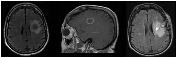
Left image: axial T2 FLAIR showing 2 cm × 2.3 cm left frontal mass with substantial edema. Middle image: sagittal T1 post-contrast image showing contrast enhancement in a “ring-like” pattern. Right image: axial T2 FLAIR showing residual changes consistent with partial resection of the previous ring-enhancing left frontal mass.
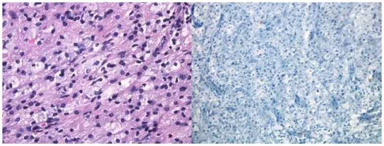
Pathological specimen of the left frontal lobe lesion. Left image: H&E section showing white matter with reactive gliosis (reactive astrocytes with abundant eosinophilic cytoplasm). Right image: Luxol fast blue showing absence of myelin.
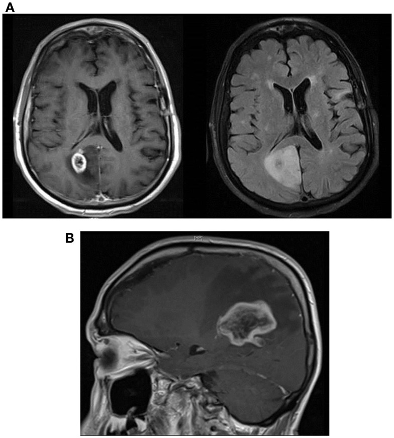
(A) Sagittal T1 post-contrast showing large area of right occipitoparietal lesion with a ring-like enhancing pattern (left image) and axial T2 FLAIR MRI showing substantial peri-lesional edema (right image). (B) Sagittal T1 post-contrast MRI showing marked increase in the size of the right parietal lobe lesion with surrounding local edema and enhancement.
Similar articles
-
Tumefactive demyelination: an approach to diagnosis and management.J Neurol Neurosurg Psychiatry. 2013 Sep;84(9):1047-53. doi: 10.1136/jnnp-2012-304498. Epub 2013 Jan 19. J Neurol Neurosurg Psychiatry. 2013. PMID: 23334629 Review.
-
Contralateral recurrence of tumefactive demyelination.Neuroradiol J. 2015 Oct;28(5):493-7. doi: 10.1177/1971400915609798. Epub 2015 Oct 1. Neuroradiol J. 2015. PMID: 26427896 Free PMC article.
-
Relapsing Tumefactive Demyelination: A Case Report.Acta Med Acad. 2018 Nov;47(2):193-198. doi: 10.5644/ama2006-124.231. Acta Med Acad. 2018. PMID: 30585071
-
Tumefactive Multiple Sclerosis Variants: Report of Two Cases of Schilder and Balo Diseases.Iran J Child Neurol. 2017 Spring;11(2):69-77. Iran J Child Neurol. 2017. PMID: 28698732 Free PMC article.
-
Retinal vasculopathy with cerebral leukoencephalopathy (RVCL): A rare mimic of tumefactive MS.Neurology. 2018 Oct 9;91(15):e1423-e1428. doi: 10.1212/WNL.0000000000006329. Epub 2018 Sep 7. Neurology. 2018. PMID: 30194247 Review.
Cited by
-
Clinical and imaging correlation in patients with pathologically confirmed tumefactive demyelinating lesions.J Neurol Sci. 2017 Oct 15;381:83-87. doi: 10.1016/j.jns.2017.08.015. Epub 2017 Aug 10. J Neurol Sci. 2017. PMID: 28991721 Free PMC article.
-
A Rare and Challenging Presentation of Acute Hemorrhagic Leukoencephalitis With Tumefactive Demyelinating Lesions in a 41-Year-Old Male.Cureus. 2024 Apr 15;16(4):e58282. doi: 10.7759/cureus.58282. eCollection 2024 Apr. Cureus. 2024. PMID: 38752096 Free PMC article.
-
Tumefactive Demyelinating Lesions in Multiple Sclerosis and Associated Disorders.Curr Neurol Neurosci Rep. 2016 Mar;16(3):26. doi: 10.1007/s11910-016-0626-9. Curr Neurol Neurosci Rep. 2016. PMID: 26847090 Review.
-
A challenging case of concurrent multiple sclerosis and anaplastic astrocytoma.Surg Neurol Int. 2019 Aug 23;10:166. doi: 10.25259/SNI_176_2019. eCollection 2019. Surg Neurol Int. 2019. PMID: 31583163 Free PMC article.
-
A challenging diagnosis of late-onset tumefactive multiple sclerosis associated to cervicodorsal syringomyelia: doubtful CT, MRI, and bioptic findings: Case report and literature review.Medicine (Baltimore). 2016 Sep;95(36):e4585. doi: 10.1097/MD.0000000000004585. Medicine (Baltimore). 2016. PMID: 27603348 Free PMC article. Review.
References
-
- Bosch G. Ein Fall von primarem Melanosarkom des Zentralnervensystemsbei multipler sklerose. Zeit Med (1912) 33:917–22.
-
- Shuangshoti S, Hjardermaal GM, Ahmad Y, Arden JL, Herman MM. Concurrence of multiple sclerosis and intracranial glioma. Report of a case and review of the literature. Clin Neuropathol (2003) 22:304–8. - PubMed
LinkOut - more resources
Full Text Sources
Other Literature Sources