

Anterior Sacral Meningocele Masquerading as an Ovarian Cyst: A Rare Clinical Presentation Associated with Marfan Syndrome
- PMID: 26236457
- PMCID: PMC4500879
- DOI: 10.4081/cp.2015.752
Anterior Sacral Meningocele Masquerading as an Ovarian Cyst: A Rare Clinical Presentation Associated with Marfan Syndrome
Abstract
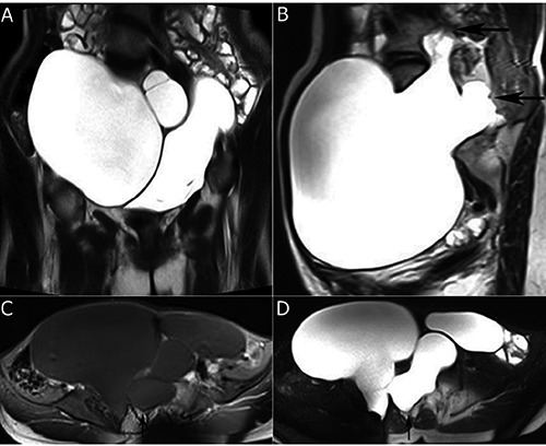
Anterior sacral meningocele is a very rare clinical entity characterized by herniation of a meningeal sac through a sacrococcygeal defect. We report a case of a 20-year old female with Marfan syndrome who presented with abdominal distention that was misdiagnosed as an ovarian cyst on pelvic ultrasound. Pelvic magnetic resonance (MR) imaging showed large, well-defined multiloculated intrasacral and presacral cysts communicating via two separate broad necks and extending through defects in anterior aspect of sacral vertebrae. This case emphasizes that anterior sacral meningocele should be considered in the differential diagnosis of cases with pelvic cysts particularly in patients with underlying connective tissue disorders. Because severe neurologic complications or even death may occur without proper preoperative planning in such cases, MR imaging should always be performed for evaluation and characterization of pelvis cystic lesions.
Keywords: Abdominal distension; Marfan syndrome; anterior sacral meningocele; ovarian cyst.
Conflict of interest statement
Conflict of interest: the authors declare no potential conflict of interest.
Figures
Similar articles
-
Anterior sacral meningocele simulating ovarian cyst.J Clin Ultrasound. 2006 Jun;34(5):244-6. doi: 10.1002/jcu.20198. J Clin Ultrasound. 2006. PMID: 16673368
-
Closure of a giant anterior sacral meningocele with an omental flap in a patient with Marfan syndrome: case report.J Neurosurg Spine. 2018 Aug;29(2):182-186. doi: 10.3171/2018.1.SPINE171303. Epub 2018 May 25. J Neurosurg Spine. 2018. PMID: 29799321
-
Anterior sacral meningocele mimicking ovarian cyst: a case report.Med Ultrason. 2013 Mar;15(1):67-70. doi: 10.11152/mu.2013.2066.151.avp1asm2. Med Ultrason. 2013. PMID: 23486628
-
Anterior sacral meningocele and Marfan syndrome: a review.Acta Chir Belg. 1993 Jan-Feb;93(1):1-7. Acta Chir Belg. 1993. PMID: 8470436 Review.
-
Holocord syringomyelia secondary to tethered spinal cord associated with anterior sacral meningocele and tailgut cyst: case report and review of literature.Childs Nerv Syst. 2014 Jun;30(6):1141-6. doi: 10.1007/s00381-014-2379-6. Epub 2014 Feb 22. Childs Nerv Syst. 2014. PMID: 24562417 Review.
Cited by
-
Atypical presentation of currarino syndrome: A case report.Int J Surg Case Rep. 2019;57:102-105. doi: 10.1016/j.ijscr.2019.02.047. Epub 2019 Mar 18. Int J Surg Case Rep. 2019. PMID: 30933899 Free PMC article.
-
Individualized management of giant anterior meningoceles-case series.Medicine (Baltimore). 2020 Apr;99(14):e19631. doi: 10.1097/MD.0000000000019631. Medicine (Baltimore). 2020. PMID: 32243391 Free PMC article.
-
Anterior sacral meningocele repair assisted by intraoperative intrathecal fluorescence and 3D printing model: illustrative case.J Neurosurg Case Lessons. 2021 May 17;1(20):CASE20159. doi: 10.3171/CASE20159. eCollection 2021 May 17. J Neurosurg Case Lessons. 2021. PMID: 35855020 Free PMC article.
-
Anterior sacral meningocele presenting as intracystic bleeding.Eur Spine J. 2018 Jul;27(Suppl 3):276-280. doi: 10.1007/s00586-017-5128-1. Epub 2017 May 18. Eur Spine J. 2018. PMID: 28523383
-
Transvaginal ultrasound-guided aspiration of an anterior sacral meningocele masquerading as a hydrosalpinx, resulting in abscess formation.BJR Case Rep. 2016 Sep 3;3(1):20160037. doi: 10.1259/bjrcr.20160037. eCollection 2017. BJR Case Rep. 2016. PMID: 30363340 Free PMC article.
References
-
- Wilkins RH. Lateral and anterior spinal meningoceles. Wilkins RH, Regachary SS, Neurosurgery, 2nd ed New York, NY: McGraw-Hill; 1996. pp 3521-5.
-
- Villarejo F, Scavone C, Blazquez MG, et al. Anterior sacral meningocele: review of the literature. Surg Neurol 1983;19:57-71. - PubMed
-
- Chen CP. Syndromes, disorders and maternal risk factors associated with neural tube defects (II). Taiwan J Obstet Gynecol 2008;47:10-7. - PubMed
-
- Shedid D, Roger EP, Benzel EC. Presacral meningocele: diagnosis and treatment. Semin Spine Surg 2006;18:161-7.
-
- Nallamshetty L, Ahn NU, Ahn UM, et al. Dural ectasia and back pain review of the literature and case report. J Spinal Disord Tech 2002;15:326-9. - PubMed
Publication types
LinkOut - more resources
Full Text Sources
Other Literature Sources