

Rare Presentation of Gall Bladder Tuberculosis in a Non Immuno-Compromised Patient
- PMID: 26236458
- PMCID: PMC4500880
- DOI: 10.4081/cp.2015.754
Rare Presentation of Gall Bladder Tuberculosis in a Non Immuno-Compromised Patient
Abstract
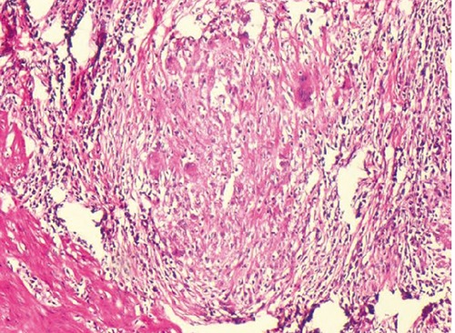
The gall bladder is least common intra-abdominal organ to be involved by tuberculosis. It is either part of systemic miliary tuberculosis or abdominal tuberculosis. Isolated gall bladder tuberculosis is even rarer, can presents either as calculus or acalculus cholecystitis. Gall bladder tuberculosis presenting as a localized perforation with a sinus formation into anterior abdominal wall is unreported complication in a non immuno-compromised person. A 48-year old female presented with a gradually increasing swelling in right hypochondrium. Abdominal ultrasound showed superficial collection over right hypochondrium with intraperitoneal extension. Computed tomography showed localized gall bladder perforation with extension to the abdominal wall. Patient underwent emergency exploration and cholecystectomy with excision of sinus tract and drainage of abdominal wall abscess. Histopathological examination showed granulomatous cholecystitis suggestive of tuberculosis of gall bladder with extension into the sinus tract. She had an uneventful recovery and was treated with 6-month antitubercular therapy after surgery.
Keywords: Gall bladder tuberculosis; gall bladder perforation.
Conflict of interest statement
Conflict of interest: the authors declare no potential conflict of interest.
Figures


Similar articles
-
Spontaneous perforation of acalculous gall bladder presenting as acute abdomen.J Emerg Med. 2012 Oct;43(4):637-40. doi: 10.1016/j.jemermed.2010.04.031. Epub 2010 Jun 26. J Emerg Med. 2012. PMID: 20580518
-
Hepatic tuberculosis--a case report.J Indian Med Assoc. 2002 Aug;100(8):522-3. J Indian Med Assoc. 2002. PMID: 12675187
-
Spontaneous Cholecystocutaneous Fistula: A rare clinical Entity.Pol Przegl Chir. 2019 Dec 9;92(5):1-5. doi: 10.5604/01.3001.0013.6277. Pol Przegl Chir. 2019. PMID: 32945267
-
Management of gall bladder perforation evaluation on ultrasonography: report of six rare cases with review of literature.J Med Life. 2011 Nov 14;4(4):364-71. Epub 2011 Nov 24. J Med Life. 2011. PMID: 22514568 Free PMC article. Review.
-
Biliary ascariasis presenting with gangrenous perforation of the gall bladder: report of a case and brief review of literature.Trop Doct. 2018 Jul;48(3):242-245. doi: 10.1177/0049475518768103. Epub 2018 Apr 13. Trop Doct. 2018. PMID: 29649951 Review.
Cited by
-
Granulomatous Lithiasic Cholecystitis in Sarcoidosis.Clin Pract. 2016 Mar 31;6(1):811. doi: 10.4081/cp.2016.811. eCollection 2016 Mar 25. Clin Pract. 2016. PMID: 27162601 Free PMC article.
-
Suspected large gall bladder mucocele extending from right hypochondrium up to right iliac region turned out to be gall bladder perforation in a patient with schizophrenia: A rare case report and literature review.Clin Case Rep. 2024 Aug 15;12(8):e9284. doi: 10.1002/ccr3.9284. eCollection 2024 Aug. Clin Case Rep. 2024. PMID: 39156201 Free PMC article.
References
-
- Rejab H, Guirat A, Ellouze S, et al. Primitive gallbladder tuberculosis: a case report with review of the literature. Ann Ital Chir 2013;84:1-3. - PubMed
-
- Sharma S, Bansal R, Agrawal N, et al. Tuberculosis of the gall bladder clinically mimicking carcinoma - a case report. J Indian Med Assoc 2012;110:402-3. - PubMed
-
- Ryu MJ, Jeon TJ, Park JY, et al. A case of gallbladder tuberculosis diagnosed by positive tuberculosis-polymerase chain reaction. Korean J Gastroenterol 2014;63:51-5. - PubMed
Publication types
LinkOut - more resources
Full Text Sources
Other Literature Sources