. 2015:2015:576743.
doi: 10.1155/2015/576743. Epub 2015 Jul 5.

Uterine Intravenous Leiomyomatosis with Cardiac Extension: Radiologic Assessment with Surgical and Pathologic Correlation

Affiliations

Uterine Intravenous Leiomyomatosis with Cardiac Extension: Radiologic Assessment with Surgical and Pathologic Correlation

Go Nakai et al. Case Rep Obstet Gynecol. 2015.

Abstract

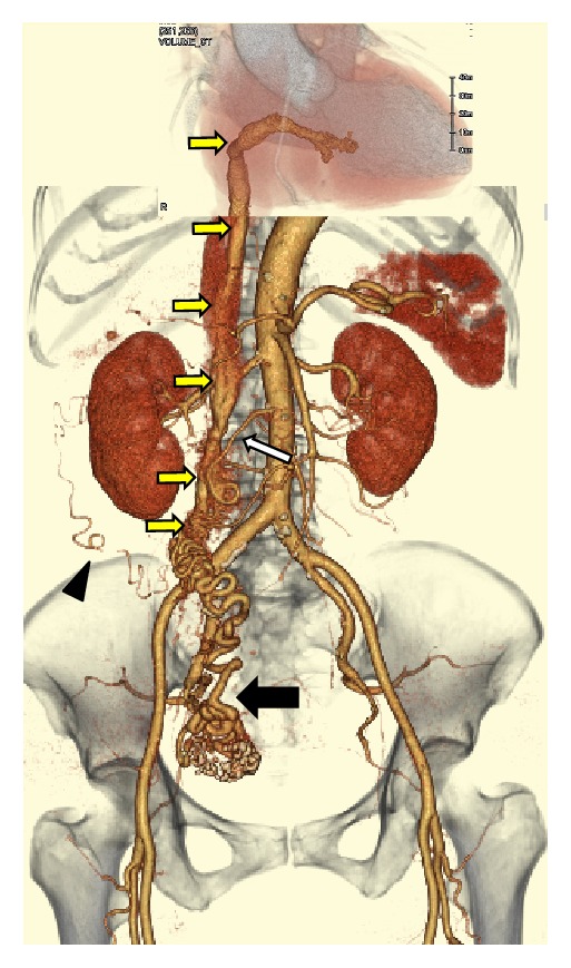
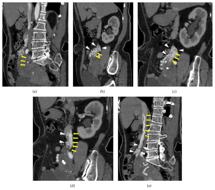
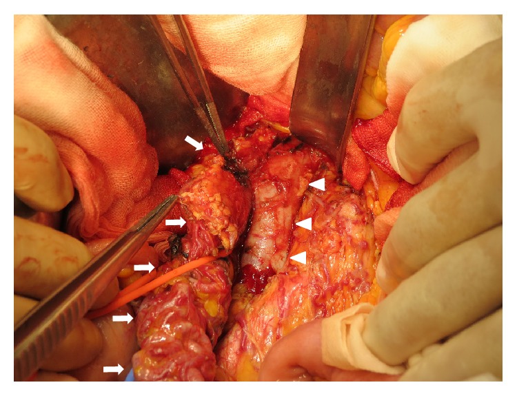
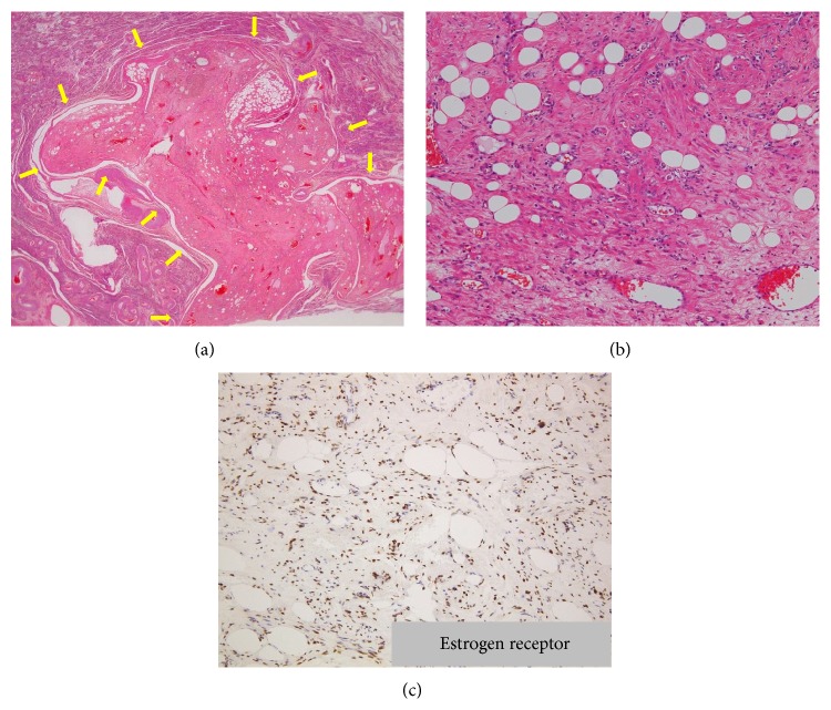
We present the computed tomography (CT) and magnetic resonance (MR) imaging findings of a 71-year-old woman with a cardiac extension of intravenous leiomyoma (IVL) that arose from the uterus, extended to the inferior vena cava (IVC), and reached the right ventricle through the right ovarian vein. Radiologic-pathologic correlation showed that the intravascular cord-like mass originating from the IVC and extending to the right ventricle was composed of degenerated smooth muscle cells with a number of large vessels that were regarded as arteries; moreover, the arteries within the cord-like mass appeared to be looping internally. Given the disappearance of the right ovarian venous wall around the IVL pathologically, extracting the tumor from the ovarian vein during an operation is considered to be impossible retrospectively. Also it was difficult to identify even the intravenous extension of the uterine leiomyoma histopathologically. Therefore, contrast-enhanced CT, in particular arterial phase imaging, provided important information that revealed the mass, range, and path of the lesion, ensuring that an appropriate operative plan could be drawn up and the tumor completely excised.


Figures

Figure 1
Figure 1
Three-dimensional arterial phase volume-rendered CT image indicated dilation of the right uterine artery (black arrow). Several small arteries originating from the right uterine artery were considered to be tumor arteries because they extended to the IVC through the right ovarian vein and then extended into the right atrium and the right ventricle (yellow arrows). A plexus of small arteries which comprised complex vascular formations originating from the right ovarian artery (white arrow), the right subcostal artery (arrowhead), and the right uterine artery surrounded tumor arteries along the route of the right ovarian vein.
Figure 2
Figure 2
(a) Multiplanar reconstruction (MPR) images along the tumor arteries allowed for identification of the origin of the intravascular component (arrows) arising from the uterine tumor (T). ((b)–(e)) A plexus of small arteries, which comprised complex vascular formations (arrowheads) originating from the right ovarian artery, the right subcostal artery, and the right uterine artery, surrounded tumor arteries (arrows) along the route of the right ovarian vein and caused the diameter of tumor arteries to expand by communicating with each other at many locations.
Figure 3
Figure 3
On MR images, the uterine tumor, approximately 13 × 7 × 6 cm in size, extensively involved the myometrium as a poorly demarcated diffuse lesion. (a) Axial T2-weighted image showed the lesion involving a few hypointense nodules (arrow) as having inhomogeneous high signal intensity (SI). (b) Axial T1-weighted image showed the lesion with a number of hyperintense tiny foci (arrow) as having low SI similar to that of the myometrium. (c) On the axial fat-suppressed T1-weighted image, the hyperintense foci within the lesion on T1-weighted images were suppressed (arrow), reflecting a lipomatous tumor. (d) On the contrast-enhanced fat-suppressed T1-weighted image, the lesion demonstrated strong contrast enhancement but contained several poorly demarcated hypointense areas, reflecting degenerative or necrotic changes. ((e), (f)) On diffusion-weighted images (e) and the ADC map (f), the lesion showed slightly high SI.
Figure 4
Figure 4
The right infundibulopelvic ligament including intravenous leiomyoma. The right infundibulopelvic (IP) ligament (arrows) including intravenous leiomyoma that arose from the uterus formed a connection to IVC (arrowheads) along the route of the right ovarian vein. A plexus of small arteries which were detected on arterial phase CT (Figures 1 and 2) was seen on the surface of the IP ligament.
Figure 5
Figure 5
(a) HE staining showed the well-circumscribed uterine lesion protruding into the uterine myometrium (arrows). (b) The uterine lesion was composed of variable amounts of smooth muscle and fat cells with hydropic and hyaline degeneration, accompanied by numerous blood vessels. (c) On immunohistochemical staining, the nuclei of smooth muscle cells in the tumor were positive for estrogen receptor (ER) and progesterone receptor (PR) (not shown).
Figure 6
Figure 6
Radiologic-pathologic correlation. (A) The level of the origin of the extrauterine lesion. (A-a) AC-CT image revealed that the origin of the continuous mass extending into the IVC was a small nodule (arrow) on the surface of the uterine tumor (arrowheads); this was determined by referring to MPR images (Figure 2(a) arrows). (A-b) On a photomicrograph at the same level as the AC-CT image, the nodule could be identified as a small nodule of lipoleiomyoma (arrows) on the surface of the uterine lipoleiomyoma (arrowheads). (A-c) No vein wall structure was found around the nodule. (B) The level of the right ovarian vein. (B-a) AC-CT indicated the presence of plexiform arteries along the route of the right ovarian vein (arrow). (B-b) A photomicrograph at the same level showed a large number of vessels (arrowheads) surrounding a bundle of vessels with a small amount of smooth muscle cells (arrow). There was no vein wall structure around smooth muscle cells and hematoxylin-eosin (HE) staining showed no evidence of the tumor component. (B-c) The nuclei of smooth muscle cells stained positive for ER and PR (not shown), which indicated that they were a continuous part of the uterine tumor, but there was no evidence of an intravenous component. (C) The level of the IVC. (C-a) The intracardiac and intracaval components of the tumor appeared as a cord-like mass composed of just a bundle of arteries on the AC-CT image (arrow). (C-b) AC-CT image showed three arteries within the tumor (arrows). (C-c) Histologic examination revealed those components were composed of degenerated smooth muscle cells including several large vessels (arrows). (C-d) The smooth muscle cells stained positive for ER and PR (not shown).

References

    1. Liu B., Liu C., Guan H., et al. Intravenous leiomyomatosis with inferior vena cava and heart extension. Journal of Vascular Surgery. 2009;50(4):897–902. doi: 10.1016/j.jvs.2009.04.037. - DOI - PubMed
    1. Du J., Zhao X., Guo D., Li H., Sun B. Intravenous leiomyomatosis of the uterus: a clinicopathologic study of 18 cases, with emphasis on early diagnosis and appropriate treatment strategies. Human Pathology. 2011;42(9):1240–1246. doi: 10.1016/j.humpath.2010.10.015. - DOI - PubMed
    1. Norris H. J., Parmley T. Mesenchymal tumors of the uterus. V. Intravenous leiomyomatosis. A clinical and pathologic study of 14 cases. Cancer. 1975;36(6):2164–2178. doi: 10.1002/cncr.2820360935. - DOI - PubMed
    1. Tierney W. M., Ehrlich C. E., Bailey J. C., King R. D., Roth L. M., Wann L. S. Intravenous leiomyomatosis of the uterus with extension into the heart. American Journal of Medicine. 1980;69(3):471–475. doi: 10.1016/0002-9343(80)90022-4. - DOI - PubMed
    1. Quade B. J., Dal Cin P., Neskey D. M., Weremowicz S., Morton C. C. Intravenous leiomyomatosis: molecular and cytogenetic analysis of a case. Modern Pathology. 2002;15(3):351–356. doi: 10.1038/modpathol.3880529. - DOI - PubMed

LinkOut - more resources