

Case Reports
doi: 10.1016/j.rmcr.2015.03.002.
eCollection 2015.
Systemic-to-pulmonary venous shunt in a patient with non-Hodgkin lymphoma: A case report and review of the literature
Affiliations
- PMID: 26236590
- PMCID: PMC4501459
- DOI: 10.1016/j.rmcr.2015.03.002
Item in Clipboard
Case Reports
Systemic-to-pulmonary venous shunt in a patient with non-Hodgkin lymphoma: A case report and review of the literature
Respir Med Case Rep.
.
Abstract
We describe a case of a systemic-to-pulmonary venous shunt secondary to superior vena cava obstruction in a patient with newly diagnosed non-Hodgkin lymphoma. This rare condition manifested with symptoms of dyspnea and hypoxemia that were out of proportion to the pleural effusion diagnosed on chest imaging. Standard treatment of such rare collateral plexuses is observation. However, it is important for clinicians to be cognizant that in rare cases such plexuses can lead to right-to-left shunt complications such as embolism.
Keywords: Non-Hodgkin lymphoma; Right-to-left shunt; Shunt; Superior vena cava; Systemic to pulmonary venous.
Figures
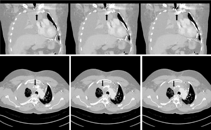
Transverse (top) and coronal (bottom) CT scan images obtained with contrast administered through a left antecubital intravenous access, showing obstruction of the SVC by a mediastinal mass (arrow).

Transverse (top) and coronal (bottom) views of CT scan images obtained with contrast administered through a right antecubital intravenous access, showing pleural enhancement (top, arrows) and bridging veins connecting intercostal and new chest wall collaterals draining into the pulmonary vein (bottom, arrows).
References
-
- Lewis M.A., Hendrickson A.W., Moynihan T.J. Oncologic emergencies: pathophysiology, presentation, diagnosis, and treatment. CA Cancer J Clin. 2011;61(5):287–314. 21858793. - PubMed
-
- Ho H.T., Horowitz A.L., Ho A.C. Systemic to pulmonary venous communication (right-to-left shunt) in superior vena cava obstruction demonstrated by spiral CT. Br J Radiol. 1999;72(859):712–713. - PubMed
-
- Kapur S., Paik E., Rezaei A., Vu D.N. Where there is blood, there is a way: unusual collateral vessels in superior and inferior vena cava obstruction. Radiographics. 2010;30(1):67–78. - PubMed
-
- Kim H.C., Chung J.W., Park S.H. Systemic-to-pulmonary venous shunt in superior vena cava obstruction: depiction on computed tomography venography. Acta Radiol. 2004;45(3):269–274. - PubMed
-
- Grayet D., Ghaye B., Szapiro D., Dondelinger R.F. Systemic-to-pulmonary venous shunt in superior vena cava obstruction revealed on dynamic helical CT. AJR Am J Roentgenol. 2001;176(1):211–213. - PubMed
Publication types
LinkOut - more resources
Full Text Sources
Other Literature Sources
