Review
. 2015 Jul 23;2(4):e135.
doi: 10.1212/NXI.0000000000000135. eCollection 2015 Aug.

Acute optic neuritis: Unmet clinical needs and model for new therapies

Affiliations
Review

Acute optic neuritis: Unmet clinical needs and model for new therapies

Steven L Galetta et al. Neurol Neuroimmunol Neuroinflamm. .

Abstract

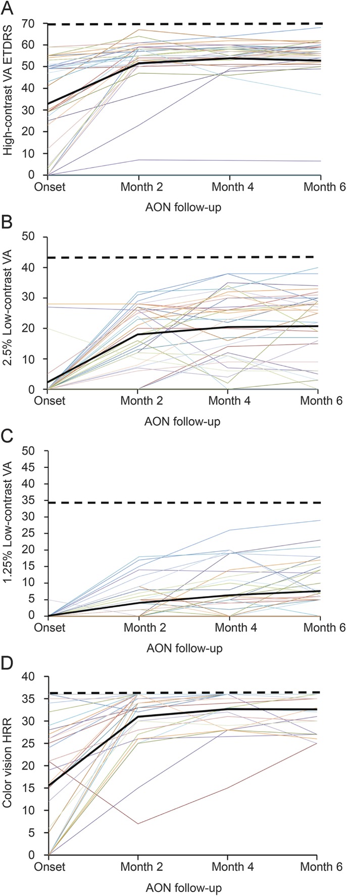
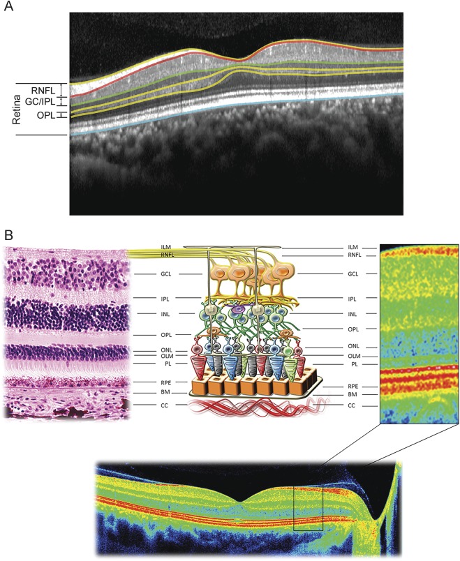
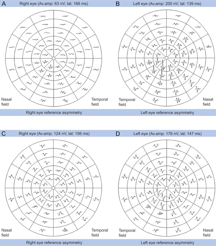
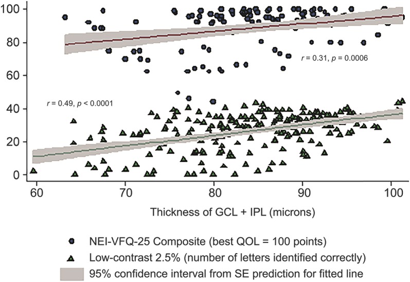
Idiopathic demyelinating optic neuritis (ON) most commonly presents as acute unilateral vision loss and eye pain and is frequently associated with multiple sclerosis. Although emphasis is often placed on the good recovery of high-contrast visual acuity, persistent deficits are frequently observed in other aspects of vision, including contrast sensitivity, visual field testing, color vision, motion perception, and vision-related quality of life. Persistent and profound structural and functional changes are often revealed by imaging and electrophysiologic techniques, including optical coherence tomography, visual-evoked potentials, and nonconventional MRI. These abnormalities can impair patients' abilities to perform daily activities (e.g., driving, working) so they have important implications for patients' quality of life. In this article, we review the sequelae from ON, including clinical, structural, and functional changes and their interrelationships. The unmet needs in each of these areas are considered and the progress made toward meeting those needs is examined. Finally, we provide an overview of past and present investigational approaches for disease modification in ON.


Figures

Figure 1
Figure 1. Evolution of visual function after acute optic neuritis
Figure shows the measurement of high-contrast visual acuity (VA) using the Early Treatment Diabetic Retinopathy Study (ETDRS) charts (A), the 2.5% and 1.25% low-contrast VA using Sloan charts (B, C), and color vision using the Hardy-Rand-Rittler (HRR) pseudoisochromatic plates (D) in a cohort of 37 patients with acute optic neuritis (AON) and visual assessment at baseline (presentation) and months 2, 4, and 6 after onset (data from Gabilondo, I et al. 2015). Each colored line is data from an individual patient, the solid black line represents the mean from all patients, and the dashed black line shows the normal values for healthy individuals for binocular testing (ETDRS = 70; 2.5% low-contrast VA = 43; 1.25% low-contrast VA = 34; HRR = 36). Reprinted with permission from Elena H. Martinez-Lapiscina.
Figure 2
Figure 2. Optical coherence tomography of the human retina
A) Detailed retinal segmentation sample on spectral-domain optical coherence tomography (OCT) image. Six intraretinal layer borders can be automatically segmented. (B) Correlation of anatomy with OCT for the human retina. On the left is a hematoxylin & eosin stain of the human retina, in the center is a schematic representation of the cell composition of the human retina, and on the right is a magnification of the image obtained with spectral-domain OCT (bottom), with indications of the retina layers identified. Figure 2B reprinted with permission from Santiago Ortiz-Perez. BM = Bruch membrane; CC = choriocapillaris layer; GCL = ganglion cell layer; ILM = inner limiting membrane; INL = inner nuclear layer; IPL = inner plexiform layer; OLM = outer limiting membrane; ONL = outer nuclear layer; OPL = outer plexiform layer; PL = photoreceptor layer; RNFL = retinal nerve fiber layer; RPE = retinal pigment epithelium.
Figure 3
Figure 3. Multifocal visual-evoked potentials in optic neuritis
Figure shows the visual-evoked potentials (VEPs) in 52 sectors of the retina. (A, B) A case of acute optic neuritis with diffuse impairment of the VEPs in the affected eye (A) compared with the unaffected eye (B), with significant impairment of the latencies and amplitudes. (B, C) After recovery from the acute optic neuritis, the VEPs show a significant decrease of amplitude and latencies in most of the sectors of the affected eye (C) compared with the unaffected eye (D). Reprinted with permission from Ana Tercero.
Figure 4
Figure 4. Relationship of thickness of retinal layers to quality of life and low-contrast visual acuity
Scatter plot and fitted linear regression line showing relationships of ganglion cell layer plus inner plexiform layer (GCL + IPL) thickness to 25-item National Eye Institute Visual Functioning Questionnaire (NEI-VFQ-25) composite scores and low-contrast visual acuity at 2.5% level. The regression lines represent fitted values for mean GCL + IPL thickness for each value of NEI-VFQ-25 or low-contrast visual acuity; the gray shaded areas show the 95% confidence intervals from the SEs of the predictions for the fitted lines. Linear correlations were significant. QOL = quality of life. Reprinted from Ophthalmology 119(6), Walter SD et al., Ganglion cell loss in relation to visual disability in multiple sclerosis, 1250–1257, 2012, with permission from Elsevier.

References

    1. Toosy AT, Mason DF, Miller DH. Optic neuritis. Lancet Neurol 2014;13:83–99. - PubMed
    1. Beck RW, Cleary PA, Anderson MM, Jr, et al. ; Optic Neuritis Study Group. A randomized, controlled trial of corticosteroids in the treatment of acute optic neuritis. N Engl J Med 1992;326:581–588. - PubMed
    1. Cole SR, Beck RW, Moke PS, Gal RL, Long DT; Optic Neuritis Study Group. The National Eye Institute Visual Function Questionnaire: experience of the ONTT. Invest Ophthalmol Vis Sci 2000;41:1017–1021. - PubMed
    1. Beck RW, Cleary PA, Backlund JC. The course of visual recovery after optic neuritis. Experience of the Optic Neuritis Treatment Trial. Ophthalmology 1994;101:1771–1778. - PubMed
    1. Costello F, Hodge W, Pan YI, Eggenberger E, Coupland S, Kardon RH. Tracking retinal nerve fiber layer loss after optic neuritis: a prospective study using optical coherence tomography. Mult Scler 2008;14:893–905. - PubMed