. 2015 Sep;11(9):705-12.
doi: 10.1038/nchembio.1870. Epub 2015 Aug 3.

Metabolic and evolutionary origin of actin-binding polyketides from diverse organisms

Affiliations

Metabolic and evolutionary origin of actin-binding polyketides from diverse organisms

Reiko Ueoka et al. Nat Chem Biol. 2015 Sep.

Abstract

Actin-targeting macrolides comprise a large, structurally diverse group of cytotoxins isolated from remarkably dissimilar micro- and macroorganisms. In spite of their disparate origins and structures, many of these compounds bind actin at the same site and exhibit structural relationships reminiscent of modular, combinatorial drug libraries. Here we investigate biosynthesis and evolution of three compound groups: misakinolides, scytophycin-type compounds and luminaolides. For misakinolides from the sponge Theonella swinhoei WA, our data suggest production by an uncultivated 'Entotheonella' symbiont, further supporting the relevance of these bacteria as sources of bioactive polyketides and peptides in sponges. Insights into misakinolide biosynthesis permitted targeted genome mining for other members, providing a cyanobacterial luminaolide producer as the first cultivated source for this dimeric compound family. The data indicate that this polyketide family is bacteria-derived and that the unusual macrolide diversity is the result of combinatorial pathway modularity for some compounds and of convergent evolution for others.


Conflict of interest statement

Competing financial interests

The authors declare no competing financial interests.

Figures
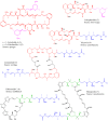
Figure 1
Figure 1
Selected structures of structurally related macrolides and their biological sources. With the exception of luminaolide, all compounds have been shown to target actin. The colored regions denote shared structural features.
Figure 2
Figure 2
Unifying model for the biosynthesis of the actin-inhibitors misakinolide A, tolytoxin, and luminaolides. a, Misakinolide biosynthetic gene cluster. b, Scytophycin and tolytoxin gene cluster. c, Luminaolide B gene cluster. The 10 kb scale bar refers to all three clusters. d, Architecture of the PKS systems and proposed macrolide biosynthesis. For luminaolides, the stereochemistry is uncertain, and the PKS is only known for luminaolide B. The first ring closure in misakinolide biosynthesis is hypothetical and might alternatively occur after polyketide assembly. Circles represent the enzymatic domains. A, adenylation domain; AT, acyltransferase; DH, dehydratase; DH*, dehydratase variant with aberrant active site motif; ER, enoylreductase; FT, formyltransferase; KR, ketoreductase; KS, ketosynthase; KS0, nonelongating KS; MT, C-methyltransferase; OMT, O-methyltransferase; PS, pyran synthase; TE, thioesterase. Solid black circled represent acyl carrier protein domains. The grey AT domain is predicted to be non-functional. Gaps between domains denote protein boundaries, dashed lines connect domains within the same protein. Black triangles point to split modules belonging to two distinct proteins for the PKS variants indicated by the labels.
Figure 3
Figure 3
Co-localization of 'Entotheonella' and misakinolide A using a combination of CARD-FISH and high resolution imaging mass spectrometry (HR-IMS). a, localization of 'Entotheonella' by CARD-FISH. Brightfield image showing representative pores from a thin section of T. swinhoei WA (left). CARD-FISH results showing 'Entotheonella' to be localized around pores of the sponge tissue (middle). Overlay of the brightfield image with the fluorescent image obtained from CARD-FISH labeling of 'Entotheonella' filaments (right). b, localization of misakinolide in the sponge tissue. Representative optical image of a T. swinhoei WA thin section used for MALDI-IMS prior to matrix application (left). False color heat map representation of the spatial distribution of misakinolide A within the sponge tissue. (middle). Overlay of the optical image and the spatial distribution of misakinolide A (right). c, Magnified section of the north-eastern part of the thin section in b. Magnified section of the north eastern part of the optical image (left). Magnified section of the north eastern part of the false color heat map representation of the spatial distribution of misakinolide A (middle). The image shows misakinolide A to be located around pores of the sponge tissue (overlapping with the observations in a). Overlay representation of the magnified optical image of the sponge tissue section and the spatial localization of misakinolide A as shown by MALDI-IMS (right).
Figure 4
Figure 4
HPLC profiles of test reactions investigating PS-catalyzed tetrahydropyran formation. i, Synthetic susbtrate 8. ii, Synthetic standard 9. iii, Assay mixture containing 8 and the expressed mis PS. iv, Assay mixture containing 8 and the boiled PS. Test substrates were racemic.
Figure 5
Figure 5
Evolutive relationships of the rhizopodin (riz), luminaolide (lum), scytophycin/tolytoxin (tto), and misakinolide (mis) PKS systems. The figure summarizes phylogenetic data of Supplementary Fig. 22-27. Numbers refer to modules carrying KSs and the corresponding domain numbering in the phylogenetic analysis (e.g., riz DH5 is the DH domain in module 5). Colors symbolize the relationship between PKS components: Blue PKS portions are closely related and likely have a common evolutionary origin. Thick green lines connect direct, late-branching neighbors in phylogenetic trees, thinner blue lines connect early-branching neighbors, black asterisks denote domains at isolated tree positions within the same clade, red asterisks label singleton KS domains in separate specificity clades. Unlabeled domains were not included in the analysis. Grey dashed lines denoted possible insertion/deletion events. Regions A and B in the mis PKS and corresponding regions in the tto and lum PKSs were likely modified by recombination at the sites marked by the red triangles.

References

    1. Allingham JS, Klenchin VA, Rayment I. Actin-targeting natural products: structures, properties and mechanisms of action. Cell Mol Life Sci. 2006;63:2119–2134. - PMC - PubMed
    1. Sasse F, Steinmetz H, Höfle G, Reichenbach H. Rhizopodin, a new compound from Myxococcus stipitatus (myxobacteria) causes formation of rhizopodia-like structures in animal cell cultures. Production, isolation, physico-chemical and biological properties. J Antibiot. 1993;46:741–748. - PubMed
    1. Ishibashi M, Moore RE, Patterson GML, Xu CF, Clardy J. Scytophycins, cytotoxic and antimycotic agents from the cyanophyte Scytonema pseudohofmanni . J Org Chem. 1986;51:5300–5306.
    1. Andrianasolo EH, et al. Isolation of swinholide A and related glycosylated derivatives from two field collections of marine cyanobacteria. Org Lett. 2005;7:1375–1378. - PubMed
    1. Sakai R, Higa T, Kashman Y. Misakinolide A, an antitumor macrolide from the marine sponge Theonella sp. Chem Lett. 1986:1499–1502.

Publication types

MeSH terms