. 2015 Aug 3;11(8):e1005042.
doi: 10.1371/journal.ppat.1005042. eCollection 2015 Aug.

Human Non-neutralizing HIV-1 Envelope Monoclonal Antibodies Limit the Number of Founder Viruses during SHIV Mucosal Infection in Rhesus Macaques

Affiliations

Human Non-neutralizing HIV-1 Envelope Monoclonal Antibodies Limit the Number of Founder Viruses during SHIV Mucosal Infection in Rhesus Macaques

Sampa Santra et al. PLoS Pathog. .

Abstract

HIV-1 mucosal transmission begins with virus or virus-infected cells moving through mucus across mucosal epithelium to infect CD4+ T cells. Although broadly neutralizing antibodies (bnAbs) are the type of HIV-1 antibodies that are most likely protective, they are not induced with current vaccine candidates. In contrast, antibodies that do not neutralize primary HIV-1 strains in the TZM-bl infection assay are readily induced by current vaccine candidates and have also been implicated as secondary correlates of decreased HIV-1 risk in the RV144 vaccine efficacy trial. Here, we have studied the capacity of anti-Env monoclonal antibodies (mAbs) against either the immunodominant region of gp41 (7B2 IgG1), the first constant region of gp120 (A32 IgG1), or the third variable loop (V3) of gp120 (CH22 IgG1) to modulate in vivo rectal mucosal transmission of a high-dose simian-human immunodeficiency virus (SHIV-BaL) in rhesus macaques. 7B2 IgG1 or A32 IgG1, each containing mutations to enhance Fc function, was administered passively to rhesus macaques but afforded no protection against productive clinical infection while the positive control antibody CH22 IgG1 prevented infection in 4 of 6 animals. Enumeration of transmitted/founder (T/F) viruses revealed that passive infusion of each of the three antibodies significantly reduced the number of T/F genomes. Thus, some antibodies that bind HIV-1 Env but fail to neutralize virus in traditional neutralization assays may limit the number of T/F viruses involved in transmission without leading to enhancement of viral infection. For one of these mAbs, gp41 mAb 7B2, we provide the first co-crystal structure in complex with a common cyclical loop motif demonstrated to be critical for infection by other retroviruses.


Conflict of interest statement

The authors have declared that no competing interests exist.

Figures
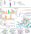
Fig 1
Fig 1. Fab and FcR binding.
(A) Linear cross-clade epitope mapping of 7B2 IgG1_AAA by peptide microarray. FcR binding (response units), on-rate (ka) and off rate (kd) by Surface Plasmon Resonance (SPR) of 7B2 IgG1_AAA. (B) Fine mapping of the 7B2 epitope within the gp41 immunodominant loop. Top Graph shows the binding response at saturation (~140 seconds after starting injection of 7B2 Fab) of each Ala-substituted gp41596-606 peptide normalized to wild type and the middle graph shows the normalized off-rate of the same peptides. Data are representative of at least two measurements on adjacent spots in the same sensor chip. Residues that are part of the 7B2 epitope are colored in orange. The Lys601Ala mutant peptide is highlighted in green since it gave a higher binding response and a decreased off-rate. Bottom graph is an example of sensogram showing 7B2 Fab binding to WT and select Alanine mutant gp41596-606 peptides that were used to generate the top and middle graphs. (C) Binding between 7B2 and gp41 peptides in standard and reducing conditions. (D) The structure of the 7B2 Fab-gp41 peptide complex shows detailed polar interactions. Hydrogen bonds between functional groups in the peptide and the heavy chain of the Fab are indicated. (E) Comparison of the gp41 ID loop from our structure (far left) against its structure obtained from NMR (middle left) [38,39] and its conformation as shown in the BG505.SOSIP.664 structure (middle right) [15] superimposed against the 7B2 paratope. A superposition of all three ID conformations (far right) highlights the conformational variability of this region.
Fig 2
Fig 2. Surface plasmon resonance of mAbs to human and rhesus FcR.
(A) 7B2 IgG1_AAA and Fab to human FcR FcgRI, FcgRIIA, FcgRIIIB and (B) rhesus macaque FcgR3A-1 and _FcgR3A-3. (C) A32 IgG1_AAA and (D) CH22 IgG1_AAA to rhesus macaque FcgR3A-1 and _FcgR3A-3.
Fig 3
Fig 3. 7B2 mAb captures infectious SHIV BaL and SF162.
SHIV BaL and SHIV SF162 virus capture by 7B2 and controls were measured by either (A) a plate–based capture assay (relative luciferase unit (RLU) on day 7 post infection shown) (B) or a column-based assay (% virus capture based on SIV gag viral RNA measurement for rVirion and RLU infectivity for iVirion percentages, respectively. The error bar is the SEM of three wells replicates. The dashed line is the positivity cutoff. The level of virion capture in the presence (red bars) and absence (blue bars) of soluble CD4 for HIV-1 SF162 (C) and HIV- BAL (D) are shown. Non-neutralizing mAb A32 and a neutralizing mAb 2G12 were used as negative and positive controls, respectively. Error bars show mean ± SEM from 3 separate experiments. (E) 7B2_AAA does not inhibit infection of rectal explants (gray) but does inhibit infectious transfer from migratory cells that emigrate from mucosal tissue (at the highest concentration (50 μg) (blue). This is reflective of CD4+ T cells being the primary targets of infection. Results are the average of two experiments.
Fig 4
Fig 4. Macrophage neutralization assays.
(A) HIV-1 Bal infected macrophages are inhibited in a dose dependent manner by 7B2-SEK and 7B2-AAA mAbs. Palivizumab did not neutralize (>100 IC90). (B) 7B2 mAb neutralization of HIV-1 SF162 (Subtype B), HIV-1 TV-1 (Subtype C), and HIV-1 Vl191. (C) 7B2_AAA displays a dose dependent inhibition of HIV infection of monocyte-derived macrophages. (D) Neutralization of BaL in peritoneal macrophages. gp41 Env specific Ab (7B2 mAb) neutralizes HIV- infection in human peritoneal macrophages. Virus input was normalized to RNA copies/mL. HIV replication was quantified by measuring the amount of luciferase in macrophage lysates.
Fig 5
Fig 5. Ability of HIV-1 Env-specific mAbs to bind HIV-1 infected cells and mediate ADCC.
(A) Mock infected primary human CD4+ T cells and (B) HIV-1 IMCBaL infected primary human CD4+ T cells were incubated with the indicated mAbs, and binding was detected by secondary staining with a FITC-conjugated anti-human IgG antibody. (C) ADCC activities of A32, CH22, and 7B2 mAbs against HIV-1IMCBaL-infected CEM.NKRCCR5 CD4+ targets cells in the presence of NK cells. Results are the average of three experiments +/- SEM.
Fig 6
Fig 6. 7B2 IgG1_AAA, A32 IgG1_AAA and CH22 IgG1_AAA mAb binding to rhesus FcR on NK cells.
(A) Schematic of experiment: Rhesus PBMC were incubated with mAb and detected with fluorescently labeled antigens specific for the mAb being tested. (B) Rhesus PBMC gated on CD16+ NK cells were analyzed for binding of an HIV-1 gp41 immunodominant region reagent. Representative data: gray curve shows binding in the absence of mAb; the black curve shows binding to a control mAb. The blue curve shifted to the right shows binding of the reagent to 7B2 mAb bound to NK cells. (C-E) Assay of PBMC from animals infused in this study. Mean fluorescence intensity of rhesus NK cells for each animal is shown (grouped here by their actual grouping in the passive infusion study). In each case, PBMC were tested using lots of mAbs used for the infusion study. Antibody-reagent pairs are as follows: C. HIV-1 gp41 immunodominant region peptide tetramer with 7B2 mAb (D) HIV-1 gp120 A244 with A32 mAb (E) HIV-1 gp120 V3 loop peptide tetramer with CH22 mAb. No differences were found between groups for each infusion set.
Fig 7
Fig 7. 7B2 IgG1_AAA, A32 IgG1_AAA and CH22 IgG1_AAA mAb concentrations in (A) plasma and (B) rectal secretions.
Concentrations of mAb were measured by a binding assay with the infused antibody as a control for calculating concentration equivalents of Ab binding to Env protein (μg/ml). Visible red blood cells in the rectal weck elutions were observed at time points post infusion for some animals.
Fig 8
Fig 8. Viral loads and CD4 T cell counts following high dose SHIV BaL rectal challenge in rhesus macaques passively infused with 7B2 IgG_AAA, A32 IgG_AAA or CH22 IgG_AAA.
(A) Plasma viral RNA levels and (B) CD4 T cell counts in 7B2 IgG_AAA and palivizumab IgG treated rhesus monkeys following challenge with SHIV-BaL. (C) Plasma viral RNA levels and (D) CD4 counts in A32 IgG_AAA mAb and control palivizumab IgG mAb passively infused rhesus monkeys following challenge with SHIV-BaL. (E) Plasma viral RNA levels and (F) CD4 Counts in rhesus monkeys following challenge with SHIV-BaL after passive administration of CH22 or CH65 IgG mAbs.
Fig 9
Fig 9. Minimum estimates of the numbers of Transmitted/Founder viruses resulting in productive clinical infection in rhesus monkeys following challenge with SHIV-BaL.
Control animals (open circles) in the 7B2 and A32 studies were treated with palivizumab, which does not bind HIV BaL. Control animals in the CH22 study were treated with CH65 IgG_AAA, an anti-influenza antibody. Treated animals (closed circles) were infused with 7B2, A32, or CH22 monoclonal antibodies, as indicated. The dashed lines represent median numbers of T/F variants among controls, while the solid lines represent median numbers of T/F variants among treated animals. Listed p-values use the Mann-Whitney rank sum t-test to determine the significance of the differences in medians between treated and control animals in each group.

References

    1. Mascola JR, Haynes BF (2013) HIV-1 neutralizing antibodies: understanding nature's pathways. Immunol Rev 254: 225–244. 10.1111/imr.12075 - DOI - PMC - PubMed
    1. Rerks-Ngarm S, Pitisuttithum P, Nitayaphan S, Kaewkungwal J, Chiu J, et al. (2009) Vaccination with ALVAC and AIDSVAX to prevent HIV-1 infection in Thailand. N Engl J Med 361: 2209–2220. 10.1056/NEJMoa0908492 - DOI - PubMed
    1. Haynes BF, Gilbert PB, McElrath MJ, Zolla-Pazner S, Tomaras GD, et al. (2012) Immune-correlates analysis of an HIV-1 vaccine efficacy trial. N Engl J Med 366: 1275–1286. 10.1056/NEJMoa1113425 - DOI - PMC - PubMed
    1. Yates NL, Liao HX, Fong Y, deCamp A, Vandergrift NA, et al. (2014) Vaccine-induced Env V1-V2 IgG3 correlates with lower HIV-1 infection risk and declines soon after vaccination. Sci Transl Med 6: 228ra239. - PMC - PubMed
    1. Tomaras GD, Ferrari G, Shen X, Alam SM, Liao HX, et al. (2013) Vaccine-induced plasma IgA specific for the C1 region of the HIV-1 envelope blocks binding and effector function of IgG. Proc Natl Acad Sci U S A 110: 9019–9024. 10.1073/pnas.1301456110 - DOI - PMC - PubMed

Publication types

MeSH terms