. 2015 Sep;33(9):980-4.
doi: 10.1038/nbt.3289. Epub 2015 Aug 3.

High-throughput determination of RNA structure by proximity ligation

Affiliations

High-throughput determination of RNA structure by proximity ligation

Vijay Ramani et al. Nat Biotechnol. 2015 Sep.

Abstract

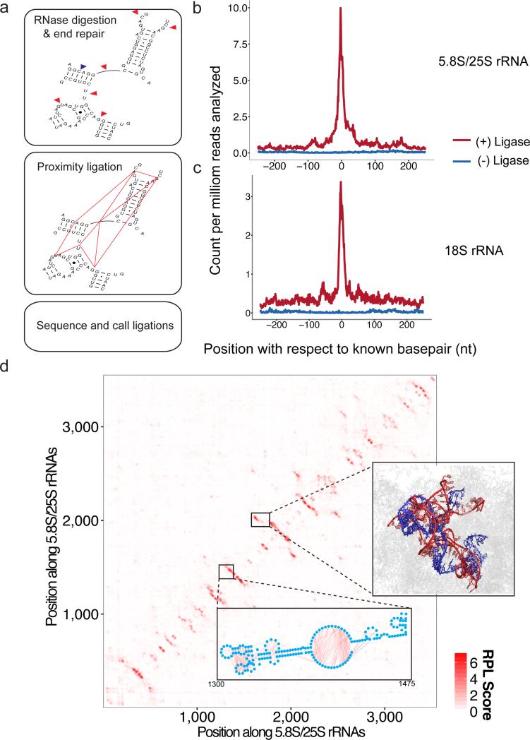
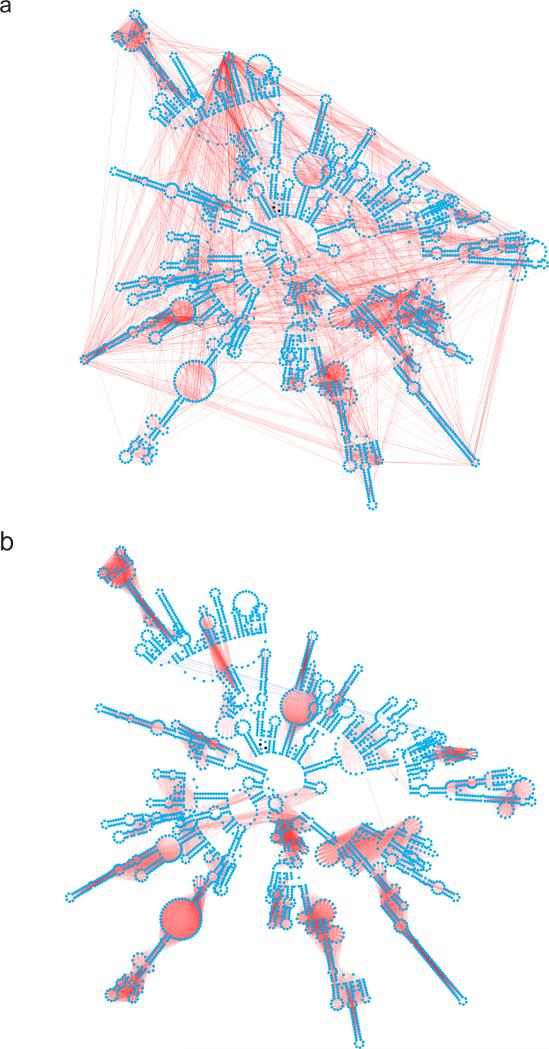
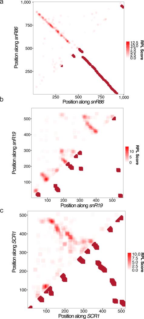
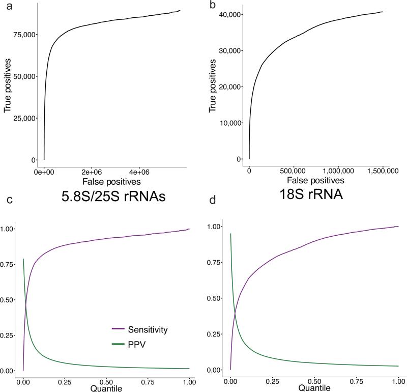
We present an unbiased method to globally resolve RNA structures through pairwise contact measurements between interacting regions. RNA proximity ligation (RPL) uses proximity ligation of native RNA followed by deep sequencing to yield chimeric reads with ligation junctions in the vicinity of structurally proximate bases. We apply RPL in both baker's yeast (Saccharomyces cerevisiae) and human cells and generate contact probability maps for ribosomal and other abundant RNAs, including yeast snoRNAs, the RNA subunit of the signal recognition particle and the yeast U2 spliceosomal RNA homolog. RPL measurements correlate with established secondary structures for these RNA molecules, including stem-loop structures and long-range pseudoknots. We anticipate that RPL will complement the current repertoire of computational and experimental approaches in enabling the high-throughput determination of secondary and tertiary RNA structures.


Figures

Figure 1
Figure 1. RNA Proximity Ligation identifies structurally proximate regions within the complex secondary structures of S. cerevisiae ribosomal RNAs. a.)
A schematic representation of the RPL method. Whole cells are spheroplasted with zymolyase and RNA is allowed to react with endogenous RNases. RNA ends are repaired in situ via T4 PNK to yield 5′-phosphate termini. Complexes are ligated overnight in the presence of T4 RNA Ligase I. Ligation products are cleaned up via acid guanidinium-phenol and subsequent DNase treatment, and subjected to Illumina TruSeq RNA-seq library preparation. These libraries are sequenced to map and count ligation junctions; b.-c.) We examined the distribution of ligation junctions as a function of distance from known base-pair partners in the 25S/5.8S rRNA and 18S rRNAs. Ligation products capture the structural proximity implied by base-pairing relationships, as evidenced by the enrichment for ligation junctions immediately near paired bases. Y-axes are shown as ligation counts per million reads analyzed. d.) Contact probability map for the eukaryotic 5.8S/25S rRNA based on RPL scores, which are calculated from the frequencies of ligation events between pairs of 21 nt windows (Methods). Lower inset: Ligation events, shown for bases 1300 to 1475 of the LSU rRNA in orange, primarily occur across digested single-stranded loops. RPL scores effectively smooth this noisy signal and are enriched for pairs of interacting regions. Plotted here are the 8,463 ligation events where both nucleotides fall within the displayed domain (compared to 17,029 ligation events where one nucleotide falls within the displayed domain and one does not, not shown). Right inset: RPL scores localize known pseudo-knots in the LSU rRNA structure, such as the interaction between bases 1727-1812 (shown in red) and bases 1941 – 2038 (shown in blue).
Figure 2
Figure 2
Smoothing of ligation junction data results in ligase-dependent signal around known stem-loop formations. a.) The 10,000 most abundant ligation pairs for the LSU rRNA (red) overlaid onto the known secondary structure (blue). While signal across stem-loops is evident, there is considerable noise. b.) Top 25,000 interacting windows based on RPL scores, which are calculated from the frequencies of ligation between pairs of 21 nt windows (Methods), for the LSU rRNA in the (+) ligase sample (red), again overlaid onto the known secondary structure (blue). Lines are drawn between the central bases of two interacting 21 nt windows. For b.), the shading of the red lines is proportional to the ligation frequency.
Figure 3
Figure 3
2D RPL contact probability maps recapitulate known and predicted non-ribosomal RNA structures. a.) Contact probability map for snR86 mirrored against interacting windows containing paired bases, based on conserved secondary structure. b.) Contact probability map for snR19 mirrored against interacting windows containing paired bases, based on conserved secondary structure. RPL signal indicating the formation of a stem-loop in bases 320-510 within the molecule is supported by MFE predictions, but not conservation. c.) Contact probability map for SCR1 mirrored against interacting windows containing paired bases, based on the known structure of SCR1. For all analyses shown here, RPL scores were calculating using a window size of 21 nt.
Figure 4
Figure 4. RPL scores demonstrate modest positive predictive value for pairs of interacting windows in RNA secondary structure. a-b.)
Plots of number of true positive interacting windows versus number of false positive interacting windows for the (a) 5.8SS/25S rRNAs and (b) 18S rRNA, at various quantile thresholds on RPL scores. This analysis shows that RPL scores have predictive value in classifying interacting regions containing at least one set of paired bases within RNA secondary structure. c-d.) Plots of the positive predictive value (green) and sensitivity (purple) of RPL-based classification of interacting regions, as a function of quantile threshold used for (c) 5.8S/25S and (d) 18S rRNAs. The quantile step size used for all analyses shown in this figure was 0.001.

Comment in

References

    1. Mortimer SA, Kidwell MA, Doudna JA. Insights into RNA structure and function from genome-wide studies. Nat. Rev. Genet. 2014;15:469–479. CrossRef Medline. - PubMed
    1. Cate JH, et al. Crystal Structure of a Group I Ribozyme Domain: Principles of RNA Packing. Science. 1996;273:1678–1685. CrossRef Medline. - PubMed
    1. Wang Y-H, Murphy FL, Cech TR, Griffith JD. Visualization of a Tertiary Structural Domain of the Tetrahymena Group I Intron by Electron Microscopy. J. Mol. Biol. 1994;236:64–71. CrossRef Medline. - PubMed
    1. Latham MP, Brown DJ, McCallum SA, Pardi A. NMR Methods for Studying the Structure and Dynamics of RNA. ChemBioChem. 2005;6:1492–1505. CrossRef Medline. - PubMed
    1. Zuker M. Mfold web server for nucleic acid folding and hybridization prediction. Nucleic Acids Res. 2003;31:3406–3415. CrossRef Medline. - PMC - PubMed

Publication types

Associated data