. 2015 Aug 26;137(33):10767-76.
doi: 10.1021/jacs.5b06644. Epub 2015 Aug 13.

A Photostable Silicon Rhodamine Platform for Optical Voltage Sensing

Affiliations

A Photostable Silicon Rhodamine Platform for Optical Voltage Sensing

Yi-Lin Huang et al. J Am Chem Soc. .

Abstract

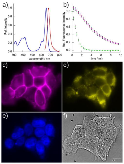
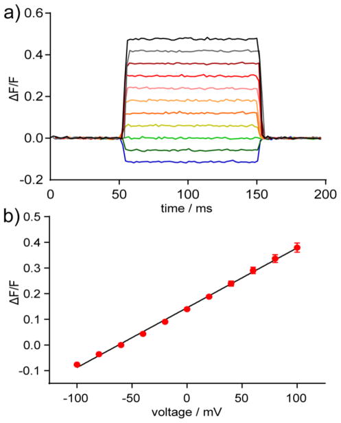
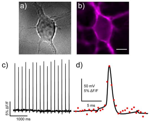
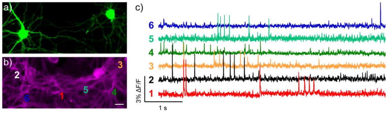
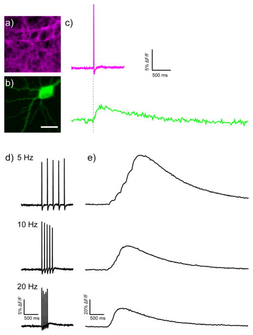
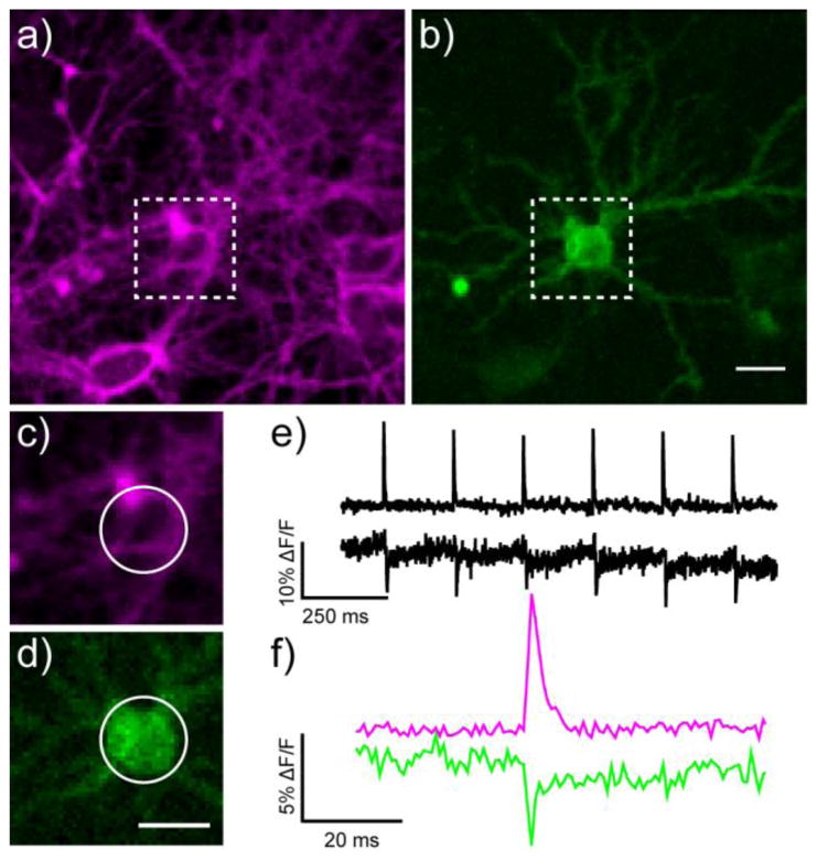
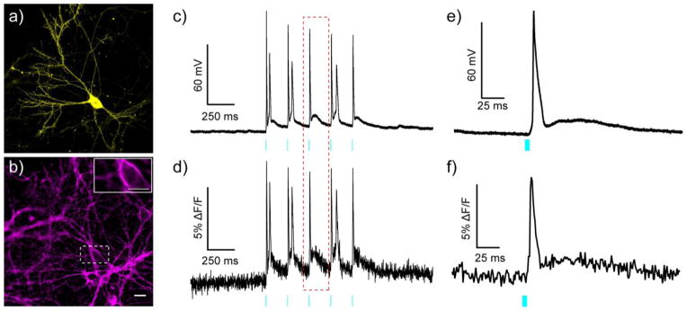
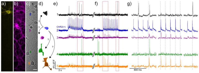
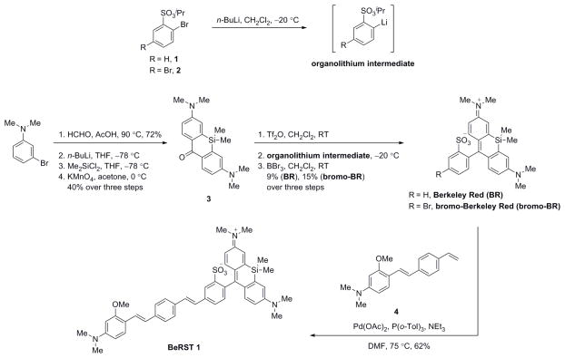
This paper describes the design and synthesis of a photostable, far-red to near-infrared (NIR) platform for optical voltage sensing. We developed a new, sulfonated silicon rhodamine fluorophore and integrated it with a phenylenevinylene molecular wire to create a Berkeley Red Sensor of Transmembrane potential, or BeRST 1 ("burst"). BeRST 1 is the first member of a class of far-red to NIR voltage sensitive dyes that make use of a photoinduced electron transfer (PeT) trigger for optical interrogation of membrane voltage. We show that BeRST 1 displays bright, membrane-localized fluorescence in living cells, high photostability, and excellent voltage sensitivity in neurons. Depolarization of the plasma membrane results in rapid fluorescence increases (24% ΔF/F per 100 mV). BeRST 1 can be used in conjunction with fluorescent stains for organelles, Ca(2+) indicators, and voltage-sensitive fluorescent proteins. In addition, the red-shifted spectral profile of BeRST 1, relative to commonly employed optogenetic actuators like ChannelRhodopsin2 (ChR2), which require blue light, enables optical electrophysiology in neurons. The high speed, sensitivity, photostability and long-wavelength fluorescence profiles of BeRST 1 make it a useful platform for the noninvasive, optical dissection of neuronal activity.


Figures

Figure 1
Figure 1
In vitro and cellular characterization of BeRST 1. Absorbance (blue) and emission (red) spectra of a) BeRST 1 in aqueous buffer (50 mM TBS, pH 7.5, 0.1% SDS). Excitation was provided at 635 nm. b) Photostability of BeRST 1 and VF2.1.Cl dyes in HEK cells. Cells were loaded with 200 nM BeRST 1 or VF2.1.Cl and then illuminated continuously for 10 min at either 631 nm (BeRST 1) or 475 nm (VF2.1.Cl) at 162 W/cm2. Images were acquired at 20 second intervals. The normalized fluorescence intensity from dye-loaded cells was plotted vs. time. Magenta circles represent BeRST 1-stained cells and green circles represent VF-2.1.Cl-stained cells. Error bars are standard error of the mean for n = 6 separate experiments. Multi-color epifluorescence imaging with BeRST 1. HEK cell were loaded with 1 μM BeRST 1, 1 μM Hoechst 33342, and 5 μM Rhodamine 123. c) BeRST 1 fluorescence localized to the cell membrane, d) Rhodamine 123 fluorescence localized to mitochondria, e) Hoechst 33342 fluorescence localized to the nucleus, and f) transmitted light DIC of HEK cells. Scale bar is 10 μm.
Figure 2
Figure 2
Voltage sensitivity of BeRST 1 in HEK cells. a) Plot of fractional change in fluorescence (ΔF/F) vs time from BeRST 1-stained HEK cells under whole-cell, voltage-clamp conditions. Cells were held at −60 mV and stepped to hyper- or de-polarizing potentials (± 100 mV) in 20 mV increments. b) Plot of ΔF/F vs. final membrane potential. Error bars are ± standard error of the mean for n = 15 cells from 3 different cultures).
Figure 3
Figure 3
Action potential (AP) visualization with BeRST 1. a) Rat hippocampal neurons were b) stained with 1 μM BeRST 1 and field stimulated to evoke action potentials that were c) recorded optically. Optical sampling frequency is 500 Hz, acquired with an EMCCD camera under 63× magnification. Scale bar is 10 μm. In a separate experiment, neurons were subjected to whole-cell current clamp and stimulated to induce action potential firing. Dual electrical and optical recording (1.8 kHz frame rate) are shown in panel (d). Black trace is the electrophysiological recording and red circles represent the optical response. A small stimulus artifact is apparent before and after the action potential in the black trace.
Figure 4
Figure 4
Spontaneous voltage imaging in GFP-labeled cells with BeRST 1. Epifluorescence images of rat hippocampal neurons expressing a) GFP and b) stained with BeRST 1. Scale bar is 20 μm. c) Optical traces of spontaneous activity in neurons from panels a and b. Numbers next to traces correspond to indicated cells in panel b. Optical sampling rate is 500 Hz.
Figure 5
Figure 5
Dual voltage and Ca2+ imaging with BeRST 1 and GCaMP6s. Epifluorescence images of a rat hippocampal neuron stained a) with BeRST 1 and expressing b) GCaMP6s. Scale bar is 20 μm. c) Sequential Ca2+ and voltage imaging during field stimulation of the neuron in panels a and b. Voltage- (upper) and Ca2+- (lower) induced changes in fluorescence. d) Voltage- (left) and e) Ca2+- (right) induced fluorescence response to trains of action potentials at the indicated frequency. Optical sample rate is 500 Hz for BeRST 1 and 40 Hz for GCaMP6s.
Figure 6
Figure 6
Two-color voltage imaging with genetically encoded voltage-fluorescent proteins and small molecule voltage-sensitive dyes. Epifluorescence images of rat hippocampal neurons a) stained with BeRST 1 and b) expressing the voltage-sensitive fluorescent protein ASAP1. Zoomed-in region for functional imaging of voltage via c) BeRST 1 staining or d) ASAP1 fluorescence. The magnified regions correspond to the white boxes in panels a and b. All scale bars are 20 μm. Trains of action potentials evoked by field stimulation at 5 Hz detected by changes in e) BeRST 1 fluorescence (upper trace) or ASAP1 fluorescence (lower trace). f) A single action potential, magnified from panel e. Magenta is BeRST 1 and green is ASAP1 fluorescence. The optical sampling rate was 1.25 kHz.
Figure 7
Figure 7
All optical electrophysiology with BeRST 1 and ChR2. Epifluorescence images of rat hippocampal neurons expressing a) YFP-ChR2 and b) stained with BeRST 1. Inset on panel b is the region inscribed by the dotted white box. The inset image is single frame used to acquire data for panels d and f. Scale bars are 20 μm (panel a/b) and 10 μm (panel b, inset). Simultaneous c) electrophysiological and d) optical recording of membrane potential changes evoked by optogenetic stimulation of the neuron expressing YFP-ChR2 and stained with BeRST 1. Cyan light (475 nm LED, 80 mW/cm2) was provided in 5 ms pulses at a rate of 5 Hz. Magnified view of e) electrophysiological and f) optical recording of the action potential highlighted in the red, dotted box from panels c and d. Optical sampling rate was 1 kHz.
Figure 8
Figure 8
Using BeRST 1 and ChR2 to perturb network activity. Cultured rat hippocampal c) neurons transfected with a) YFP-ChR2 and stained with b) BeRST 1 were stimulated with 475 nm light (80 mW/cm2, 5 ms, 5 Hz, cyan bars) during two separate 3 second periods to evoke activity in the YFP-ChR2-expressing cell. Scale bar is 20 μm. d) Schematic representation of neurons from DIC image in panel c), color-coded to match the corresponding traces in e–g). The blue ChR2(+) cell is depicted making possible connections to other neurons in the field of view. Optical records of BeRST 1 responses were acquired at 500 Hz with an sCMOS camera during e) an optical recording session and f) subsequent trial, separated by approximately 30 seconds (double hash). Numbers and colors of traces refer to specific neurons in panels a–d. Red boxes indicate areas of the trace that have been magnified for clarity in panel g). Dotted grey lines are provided in panel g) to help visually estimate the spike timing of BeRST 1-stained neurons.
Scheme 1
Scheme 1
Synthesis of Berkeley Red Sensor of Transmembrane Potential 1 (BeRST 1)

References

    1. Hamill OP, Marty A, Neher E, Sakmann B, Sigworth FJ. Pflugers Arch. 1981;391:85. - PubMed
    1. Spruston N, Jaffe DB, Williams SH, Johnston D. J Neurophysiol. 1993;70:781. - PubMed
    1. Peterka DS, Takahashi H, Yuste R. Neuron. 2011;69:9. - PMC - PubMed
    1. Scanziani M, Hausser M. Nature. 2009;461:930. - PubMed
    1. Paredes RM, Etzler JC, Watts LT, Zheng W, Lechleiter JD. Methods. 2008;46:143. - PMC - PubMed

Publication types