. 2015 Aug 4:6:7844.
doi: 10.1038/ncomms8844.

Nonsynaptic junctions on myelinating glia promote preferential myelination of electrically active axons

Affiliations

Nonsynaptic junctions on myelinating glia promote preferential myelination of electrically active axons

Hiroaki Wake et al. Nat Commun. .

Abstract

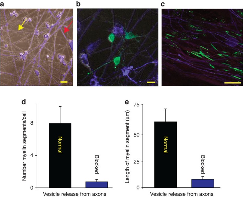
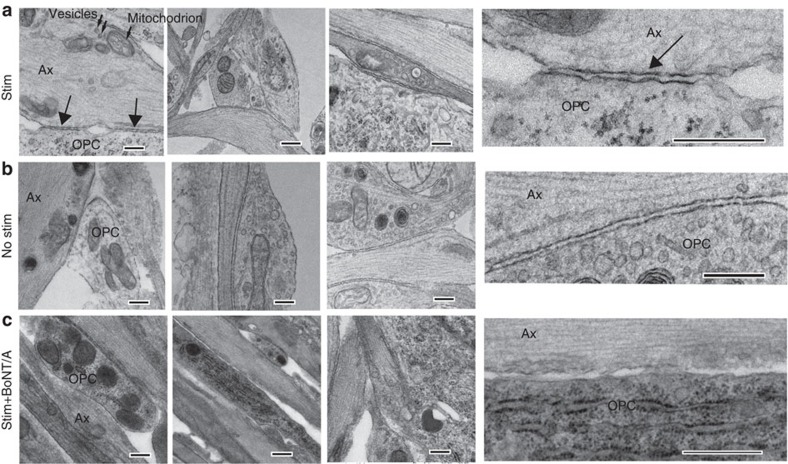
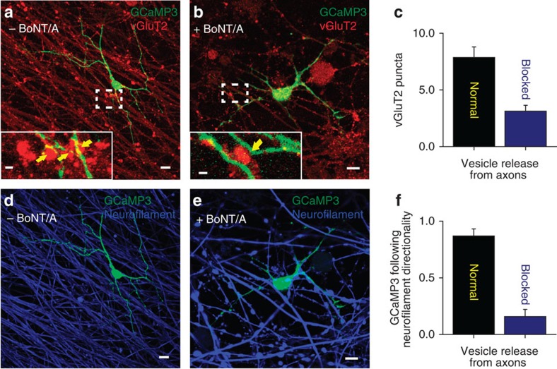
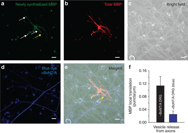
The myelin sheath on vertebrate axons is critical for neural impulse transmission, but whether electrically active axons are preferentially myelinated by glial cells, and if so, whether axo-glial synapses are involved, are long-standing questions of significance to nervous system development, plasticity and disease. Here we show using an in vitro system that oligodendrocytes preferentially myelinate electrically active axons, but synapses from axons onto myelin-forming oligodendroglial cells are not required. Instead, vesicular release at nonsynaptic axo-glial junctions induces myelination. Axons releasing neurotransmitter from vesicles that accumulate in axon varicosities induces a local rise in cytoplasmic calcium in glial cell processes at these nonsynaptic functional junctions, and this signalling stimulates local translation of myelin basic protein to initiate myelination.


Figures

Figure 1
Figure 1. Electrically active axons releasing synaptic vesicles are preferentially myelinated.
(a) DRG neurons treated with BoNT/A and stained with cell tracker (blue, see red arrow) co-cultured with normal (untreated) neurons (grey, see yellow arrow). (b) OPCs (green, GCaMP3) were added to the cultures to determine whether exocytosis of neurotransmitter from axons influenced myelination. (c) Axons were stimulated for 9 s at 10 Hz every 5 min for 10 h and cultured for 3 weeks. Myelin (green, myelin basic protein, MBP) analysed 3 weeks after co-culture formed preferentially on axons releasing synaptic vesicles (purple, neurofilament), and (d) number of myelin segments/cell were more in normal axons (P<0.001, n=7 cells from four dishes) (e) myelin segments were also longer in normal axons (P<0.001, n=9 cells from four dishes). Scale bar, 10 μm (a,b); 20 μm (c).
Figure 2
Figure 2. Myelination occurs in absence of axon-OPC AMPAR-mediated synaptic currents.
(a) Currents elicited in a recorded OPC held at −80 mV 1 day after plating by voltage steps from +20 to −110 mV. Note the presence of sodium currents (inset). (b) IV curve of sodium currents for the same OPC after leak subtraction (inset). (c) Lack of evoked synaptic currents in an OPC upon DRG axonal stimulation. There was no response in 74 cells tested (unstimulated n=17 cells, from seven dishes; pre-stimulated electrically with (n=15 cells from six dishes) or without (n=42 from 17 dishes) BoNT/A). Individual (grey, 15 sweeps) and average traces (black) are shown. Axonal stimulation time is indicated with arrowheads. (d) Bath application of the secretagogue ruthenium red (75 μM) in absence of electrical DRG axonal stimulation did not evoke any currents in the same OPC. Capacitive currents in response to a test pulse are shown (unstimulated n=10 cells from four dishes, pre-stimulated n=18 cells from seven dishes and pre-stimulated with BoNT/A n=7 cells from four dishes). (e,f) Spontaneous (e) and miniature (f) synaptic currents in an OPC recorded at 15 postnatal days in acute coronal corpus callosum slices of NG2-DsRed mice (N=2 mice). Miniature synaptic currents were recorded in 1 μM tetrodotoxin (TTX) and 75 μM ruthenium red (Rred). The mean frequency, rise and decay times of spontaneous synaptic activity are 0.51 Hz, t10–90%=332 μs and τ=1.35 ms, respectively (insets, n=7 cells from seven different brain slices). The holding potential is indicated for each trace.
Figure 3
Figure 3. Ca2+ increases in processes of OPCs at axonal varicosities.
(a) OPC processes (green, GCaMP3 transfection) form specialized functional junctions with DRG axons (blue, neurofilament), containing accumulations of synaptic vesicles containing glutamate (red, vGluT2). Scale bar, 10 μm. After live-cell calcium imaging (b), the cultures were fixed and stained by immunocytochemistry to determine whether that the calcium responses were associated with axo-glial contacts containing glutamatergic synaptic vesicles (inset in a). Note colocalization between axonal varicosity (red, vGluT2) and swellings in OPC process (green, GCaMP3). Scale bar, 2 μm. (b) Time-lapse series showing a local increase in Ca2+ in OPC-axon junctions in response to electrical stimulation of axons. A stimulus-induced, local increase in Ca2+ in the same axo-glial junction (yellow square) that is shown in a is recorded in the OPC transfected with GCaMP3. Time since stimulus onset is shown in each frame (5.5–13 s). The fluorescence intensities at the axo-glial contact (yellow square) and the OPC cell body (yellow circle) are shown. Note the local increase in fluorescence intensity at the axo-glial junction after stimulation but this is not accompanied by an increase in fluorescence intensity in the soma. Scale bar, 5 μm.
Figure 4
Figure 4. Ca2+ increase in OPC processes is mediated by glutamate and ATP released in response to action potentials in axons.
(a) Representative Ca2+ traces in OPC processes in response to electrical stimulation of axons without (black) and with (blue) BoNT/A treatment to block SNARE-dependent exocytosis. (b) Averaged Ca2+ traces in OPCs from 14 dishes are shown. Responses were inhibited by BoNT/A treatment. **P=0.001, t-test, peak amplitude, n=14 cells from 14 dishes with no BoNT/A, n=14 cells from 14 dishes with BoNT/A. (c) Number of Ca2+ responses were reduced significantly by BoNT/A treatment (blocked). The peak Ca2+ response within 200 s of axonal stimulation (10 Hz) were measured. P=0.004, t-test, n=14 cells from 14 dishes with no BoNT/A, n=14 cells from 14 dishes with BoNT/A. (d) Summary graph shows per cent inhibition of Ca2+ responses following treatment with selective blockers of glutamatergic and purinergic neurotransmitter receptors, including a combination of CNQX, DAPV, MCPG (GluR antagonists), suramin and MCPG. The results implicate both glutamate and ATP neurotransmitter signalling. P=0.3, n=6 cells from six dishes, *1P=0.03, n=4 cells from four dishes, **P=0.008, n=5 cells from five dishes, *2P=0.02, n=6 cells from six dishes, all paired t-test.
Figure 5
Figure 5. Nonsynaptic junctions between axons and OPCs are promoted by vesicular release from axons.
Transmission electron microscopy showed specialized contacts (arrows) between OPC processes (OPC) and axon varicosities (Ax) containing intracellular vesicles (small arrows in a) and mitochondrion, but no synapses were detected. Three examples for each condition are shown. Insets (right column) show these junctions at higher magnification. Such contacts were evident in cultures stimulated for 9 s at 10 Hz every 5 min for 10 h (Stim) before plating OPCs, and unstimulated cultures (b), but such contacts were not found in stimulated cultures treated with BoNT/A before adding OPCs (c). Scale bar, 1 μm.
Figure 6
Figure 6. OPC processes preferentially contact electrically active axons releasing synaptic vesicles and form nonsynaptic axo-glial junctions.
(a,b) Images showing OPCs transfected with GCaMP3 construct (green) and vGluT2 immunocytochemistry (red) to identify glutamate-containing vesicles in axons. OPCs were plated on axons either previously treated (b) or not treated (a) with BoNT/A. (a,b) Magnified views of glutamate-containing vesicles and OPC processes. Yellow arrows indicate vGluT2 stained puncta in close opposition to GCaMP3 processes. (c) The number of vGluT2 puncta >0.5 μm in diameter was reduced significantly on axons previously treated with BoNT/A (P=0.0005, t-test, n=7 cells from seven dishes with no BoNT/A, n=8 cells from eight dishes with BoNT/A). (d,e) Images showing OPC transfected with GCaMP3 construct (green) and immunocytochemistry for neurofilament (blue, an axon marker). (f) Summary graph showing that the fraction of all OPC processes (d,e) in individual OPCs forming parallel associations with axons was significantly reduced when axons were previously treated with BoNT/A (P=0.0002, t-test, n=7 cells from seven dishes with no BoNT/A, n=8 cells from eight dishes with BoNT/A). Scale bars, 10 μm (a, b,d,e); 2 μm (a,b).
Figure 7
Figure 7. Local translation of MBP occurs preferentially on OPC processes contacting electrically active axons releasing synaptic vesicles.
(a) Newly synthesized MBP (green). Axons were stimulated electrically at 10 Hz for 10 min and local translation of MBP was monitored using kikume MBP fluorescence after 40 min. White arrows indicate new MBP translation in contrast yellow arrow showing no new MBP translation. (b) Total MBP (red). (c) Bright field showing cell morphology of co-culture. (d) Axons treated previously with BoNT/A to block synaptic vesicle release (blue). (e) Combined images from ad. Differential interference contrast and fluorescence to identify axons treated (blue, yellow arrow) and not treated with BoNT/A. Yellow arrow shows axon and OPC interaction without MBP translation (notice yellow arrow in a with lack of new MBP green puncta indicating no new MBP translation). (f) Quantification shows significantly more local translation of MBP in OPC processes that were in contact with axons releasing synaptic vesicles (grey axons) (0.11±0.024 versus 0.028±0.011 puncta per micrometre length of axon not treated or previously treated with BoNT/A respectively, P<0.001, t-test, n=24 cells from non-blue, n=23 cells from blue axons, five dishes). Scale bar, 10 μm.

References

    1. Bergles D. E., Roberts J. D., Somogyi P. & Jahr C. E. Glutamatergic synapses on oligodendrocyte precursor cells in the hippocampus. Nature 405, 187–191 (2000). - PubMed
    1. Emery B. Regulation of oligodendrocyte differentiation and myelination. Science 330, 779–782 (2010). - PubMed
    1. Fields R. D. Chapter in Neuroglia 3rd edn eds Kettenmann H., Ransom B. R. 573–585Oxford Univ. Press (2013).
    1. Mangin J. M. & Gallo V. The curious case of NG2 cells: transient trend or game changer? ASN Neuro 3, e00052 (2011). - PMC - PubMed
    1. Ziskin J. L., Nishiyama A., Rubio M., Fukaya M. & Bergles D. E. Vesicular release of glutamate from unmyelinated axons in white matter. Nat. Neurosci. 10, 321–330 (2007). - PMC - PubMed

Publication types

LinkOut - more resources