. 2015 Aug 4:6:7871.
doi: 10.1038/ncomms8871.

Crystal structure, biochemical and cellular activities demonstrate separate functions of MTH1 and MTH2

Affiliations

Crystal structure, biochemical and cellular activities demonstrate separate functions of MTH1 and MTH2

Megan Carter et al. Nat Commun. .

Abstract

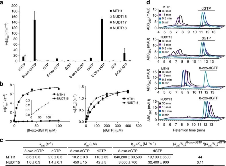
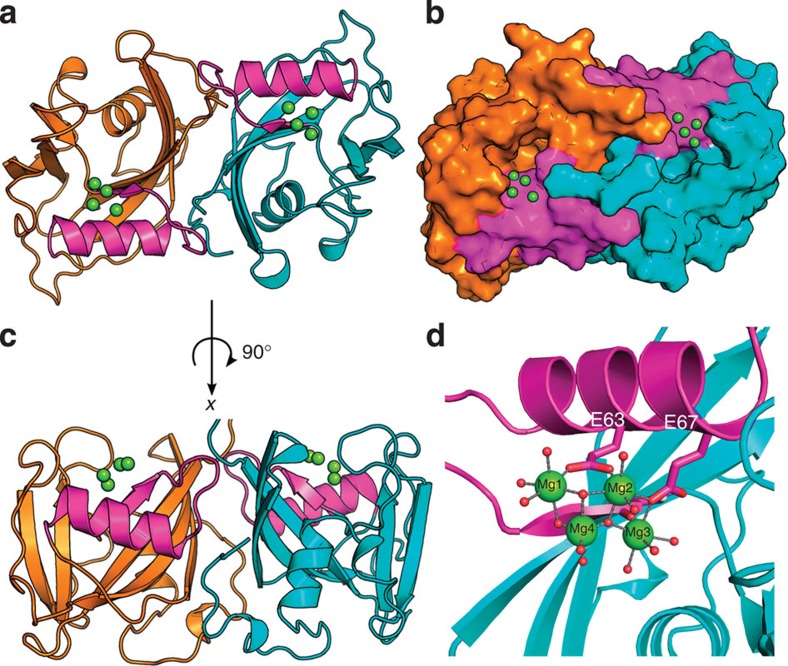
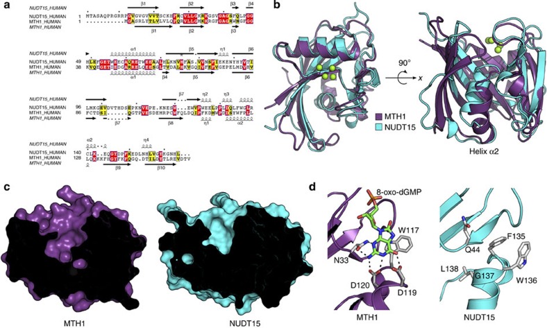
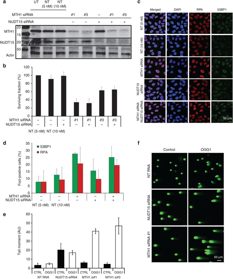
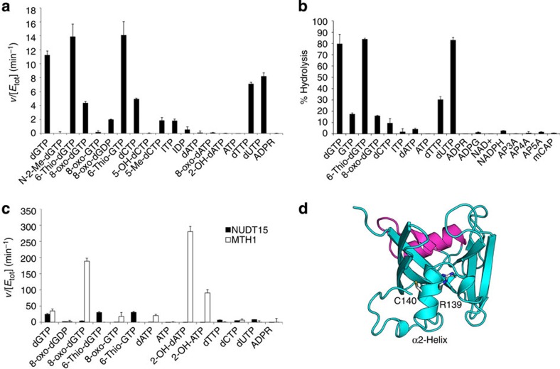
Deregulated redox metabolism in cancer leads to oxidative damage to cellular components including deoxyribonucleoside triphosphates (dNTPs). Targeting dNTP pool sanitizing enzymes, such as MTH1, is a highly promising anticancer strategy. The MTH2 protein, known as NUDT15, is described as the second human homologue of bacterial MutT with 8-oxo-dGTPase activity. We present the first NUDT15 crystal structure and demonstrate that NUDT15 prefers other nucleotide substrates over 8-oxo-dGTP. Key structural features are identified that explain different substrate preferences for NUDT15 and MTH1. We find that depletion of NUDT15 has no effect on incorporation of 8-oxo-dGTP into DNA and does not impact cancer cell survival in cell lines tested. NUDT17 and NUDT18 were also profiled and found to have far less activity than MTH1 against oxidized nucleotides. We show that NUDT15 is not a biologically relevant 8-oxo-dGTPase, and that MTH1 is the most prominent sanitizer of the cellular dNTP pool known to date.


Figures

Figure 1
Figure 1. Comparison of NUDIX protein activity with nucleotide substrates.
(a) Nucleotide substrate (50 μM) was incubated with 5–500 nM NUDIX protein depending on enzyme. Hydrolysis was monitored by detecting phosphate generated. The depicted data are representative of two independent experiments showing the same result. Data are presented as v (hydrolysed substrate (μM) per minute) per [enzyme] (μM). (b) Saturation curves and kinetic parameters of MTH1- and NUDT15-mediated hydrolysis of 8-oxo-dGTP (left) and dGTP (right). NUDT15 (8 nM) or MTH1 (0.25 nM) was incubated with 8-oxo-dGTP at concentrations ranging from 0 to 100 μM in assay buffer, and initial rates were determined in duplicate. Inset highlights NUDT15 activity on a smaller activity scale. NUDT15 (8 nM) and MTH1 (2 nM) were incubated with dGTP in assay buffer ranging from 0 to 400 μM, and initial rates were determined in duplicate. Data are presented as v (hydrolysed substrate (μM) per second) per [enzyme] (μM), and are representative of data collected from at least two independent experiments. (c) Kinetic parameters of MTH1 and NUDT15 for dGTP and 8-oxo-dGTP hydrolysis. The Michaelis–Menten equation was applied to saturation curves using the GraphPad Prism software and kinetic parameters were calculated. Data presented are average±s.d. from two independent experiments. (d) HPLC chromatograms showing the activity of NUDT15 and MTH1 against 8-oxo-dGTP and dGTP. The depicted data are representative of three independent experiments and show that MTH1 rapidly hydrolyses 8-oxo-dGTP; however, no significant activity is observed with NUDT15. Both MTH1 and NUDT15 can hydrolyse dGTP.
Figure 2
Figure 2. Crystallographic structure of dimeric NUDT15.
(a) Top view of Nudt15 dimer (chain A in cyan and chain B in orange) ribbon representation with Mg coordination, and the NUDIX box highlighted in magenta. (b) Surface representation of NUDT15 dimer looking into the putative binding pocket. (c) Side view of NUDT15 dimer. (d) Four Mg ions in coordination (green spheres) with multiple waters (red spheres), E63, E67 and carbonyl oxygen of G47 residues in the NUDIX box (magenta).
Figure 3
Figure 3. Structural comparison and sequence alignment of NUDT15 and MTH1.
(a) Sequence alignment of human NUDT15 with human MTH1 (29% overall sequence identity). The secondary structure of NUDT15 is displayed above the sequence alignment and that of MTH1 below the alignment. Sequence similarity is represented by yellow boxes, strict sequence identity in red boxes, beta sheets (β) as arrows, alpha-helices (α) and 310-helices (η) as squiggles. (b) Superimposition of MTH1 (purple) and NUDT15 (chain A, cyan) with the NUDT15 Mg2+ coordination is shown. Major structural deviation is observed in helix α2 that affects the depth of the putative binding pocket. (c) Comparison of NUDT15 and MTH1 binding pocket depth. Cut view of surface representation highlights the variation in binding pocket depth of MTH1 (purple) and Nudt15 (cyan). (d) Comparison of NUDT15 and MTH1 binding pocket composition. MTH1 helix α2 residues W117, P118, D119, D120 and N33 (grey) are involved in 8-oxo-dG (green) binding. NUDT15 helix α2 residues F135, W136, G137, L138 and Q44 (grey) form the bottom of the putative binding pocket and are unable to make the equivalent hydrogen bonding network observed in MTH1 binding to 8-oxo-dGMP.
Figure 4
Figure 4. Effect of NUDT15 knockdown on clonogenic survival, DNA damage responses and 8-oxo-dG levels in DNA.
(a) Western blot showing knockdown of MTH1 or NUDT15 in U2OS cells 96 h after transfection. (b) U2OS cells depleted of MTH1, NUDT15 or both by siRNA were seeded for clonogenic survival. Colonies were stained after 10 days with methylene blue and colonies were counted by eye; (n=2). (c) Representative immunostainings. After NUDT15, MTH1 or combination of both RPA foci, 53BP1 foci and DAPI (4,6-diamidino-2-phenylindole) were visualized using immunofluorescence. (d) Quantification of 53BP1- and RPA-positive cells (>5 foci per cell) from c; (n=2). (e) Quantification of the tail moment after MTH1 and NUDT15 knockdown; (n=2). (f) Representative pictures of comets formed after MTH1 or NUDT15 depletion by siRNA. Lysed cells were treated with OGG1 or buffer control. UT, untransfected; NT, non-targeting siRNA control. Data shown as average±s.d.
Figure 5
Figure 5. Activity of NUDT15 towards potential NUDIX family substrates.
(a) Substrate hydrolysis after 30 min incubation at 22 °C was monitored by coupling the reaction to an excess of bovine alkaline phosphatase or E. coli pyrophosphatase and measuring inorganic phosphate using malachite green reagent. Data are presented as v (hydrolysed substrate (μM) per minute) per [NUDT15] (μM). Depicted data are mean±s.d. of triplicates. Experiment was performed twice with similar results. (b) Relative activity of NUDT15 by HPLC analysis. Activity of 10 nM NUDT15 protein against a panel of potential NUDIX substrates was measured by HPLC after 30 min incubation at 37 °C. Enzymatic activity was stopped by addition of trifluoroacetic acid (5%) and samples were ran under conditions indicated in Methods section. Per cent hydrolysis was calculated by subtracting the peak area at 30 min from the area at time 0 and dividing this by the area at time 0; (n=2). (c) Substrate hydrolysis after 20 min incubation at 22 °C with NUDT15 (black bars) or MTH1 (white bars). Substrate hydrolysis was monitored by coupling the reaction to phosphatase or pyrophosphatase and measuring the presence of inorganic phosphate as described above. Experiments were performed in duplicate and data are presented as v (hydrolysed substrate (μM) per minute) per [enzyme] (μM). The experiment was repeated with similar results. (d) Mutation of NUDT15 (cyan) Arg139 to Cys is implicated in thiopurine-induced leukopaenia. Arg139 is located at the base of the helix α2 adjacent to Cys140. The NUDIX box is shown in magenta.

References

    1. Cheung-Ong K., Giaever G. & Nislow C. DNA-damaging agents in cancer chemotherapy: serendipity and chemical biology. Chem. Biol. 20, 648–659 (2013). - PubMed
    1. Bouwman P. & Jonkers J. The effects of deregulated DNA damage signalling on cancer chemotherapy response and resistance. Nat. Rev. Cancer 12, 587–598 (2012). - PubMed
    1. Fu D., Calvo J. A. & Samson L. D. Balancing repair and tolerance of DNA damage caused by alkylating agents. Nat. Rev. Cancer 12, 104–120 (2012). - PMC - PubMed
    1. Rai P. et al. Enhanced elimination of oxidized guanine nucleotides inhibits oncogenic RAS-induced DNA damage and premature senescence. Oncogene 30, 1489–1496 (2011). - PubMed
    1. Zhang Y. et al. Redox control of the survival of healthy and diseased cells. Antioxid. Redox Signal 15, 2867–2908 (2011). - PubMed

Publication types