Comparative Study
. 2015 Aug 4:10:163.
doi: 10.1186/s13014-015-0453-1.

The Focinator - a new open-source tool for high-throughput foci evaluation of DNA damage

Affiliations
Comparative Study

The Focinator - a new open-source tool for high-throughput foci evaluation of DNA damage

Sebastian Oeck et al. Radiat Oncol. .

Abstract

Background: The quantitative analysis of foci plays an important role in many cell biological methods such as counting of colonies or cells, organelles or vesicles, or the number of protein complexes. In radiation biology and molecular radiation oncology, DNA damage and DNA repair kinetics upon ionizing radiation (IR) are evaluated by counting protein clusters or accumulations of phosphorylated proteins recruited to DNA damage sites. Consistency in counting and interpretation of foci remains challenging. Many current software solutions describe instructions for time-consuming and error-prone manual analysis, provide incomplete algorithms for analysis or are expensive. Therefore, we aimed to develop a tool for costless, automated, quantitative and qualitative analysis of foci.

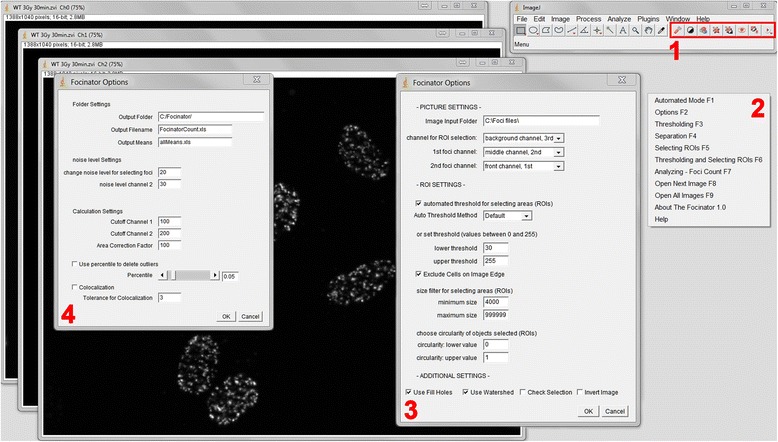
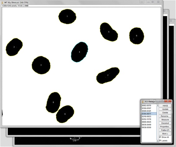
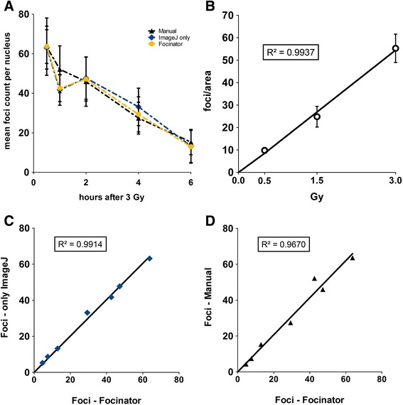
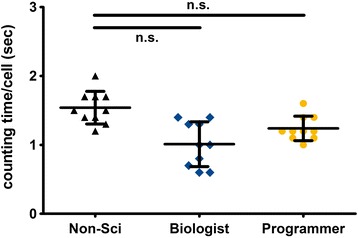
Methods: For this purpose we integrated a user-friendly interface into ImageJ and selected parameters to allow automated selection of regions of interest (ROIs) depending on their size and circularity. We added different export options and a batch analysis. The use of the Focinator was tested by analyzing γ-H2.AX foci in murine prostate adenocarcinoma cells (TRAMP-C1) at different time points after IR with 0.5 to 3 Gray (Gy). Additionally, measurements were performed by users with different backgrounds and experience.

Results: The Focinator turned out to be an easily adjustable tool for automation of foci counting. It significantly reduced the analysis time of radiation-induced DNA-damage foci. Furthermore, different user groups were able to achieve a similar counting velocity. Importantly, there was no difference in nuclei detection between the Focinator and ImageJ alone.

Conclusions: The Focinator is a costless, user-friendly tool for fast high-throughput evaluation of DNA repair foci. The macro allows improved foci evaluation regarding accuracy, reproducibility and analysis speed compared to manual analysis. As innovative option, the macro offers a combination of multichannel evaluation including colocalization analysis and the possibility to run all analyses in a batch mode.


Figures

Fig. 1
Fig. 1
The ImageJ-based interface of the Focinator offers options to adapt the evaluation parameters to distinct image characteristics. Figure 1 shows ImageJ with the Focinator macro installed as start-up macro after opening a multi-channel image. This microscope image with the file format ZVI 16-bit includes three fluorescence channels. The main window of the Focinator is implemented into the ImageJ window. It consists of a menu (2), buttons (1) and Focinator Options (3 and 4). The Focinator Options windows offer several preferences for the user to adapt the macro’s behavior to individual requirements. Picture Settings: First step is to tell the macro, the input folder and if there is a multi-channel image or more single pictures will be opened. In the second step you choose in which channel the foci have to be counted and where the ROIs should be selected. In our example, the γ-H2.AX foci are in channel number 2 (on top after opening the image). The macro will use the setting “1st foci channel = front channel” for all pictures automatically. If no second foci channel is used the setting should be changed to “inactive”. ROI Settings (3): Depending on image quality, size and magnification, it is recommended to set the threshold and the size filters for ROIs. Alternatively, the choice of automated thresholding is possible. It is possible to exclude objects that are partially outside of the image. If there are objects to exclude because they are not circular enough or too small, it is possible to exclude them via circularity filters or size filters. “Use fill holes” should be activated, if the ROI selection left holes in the cells. Overlapping ROIs (cells, nuclei) might be separated by choosing “watershed”. Regarding the batch mode “check selection” offers the possibility of stopping during the selection process. “Invert images” should be checked when working with images with light background. For the automated batch (4) mode, output directories need to be chosen to save the results. An important step of evaluation is to choose the right noise level. Noise level values can be set independently in multi-channel analysis to exclude background artifacts. By defining the cut off, foci with intensities below a certain value are deleted, which excludes background noise. The value for area correction is dependent on the mean size of the analyzed nuclei. The factor corrects the foci number divided by the individual area of each nucleus. The usage of the percentile option enables the user to delete the outliers, such as cells with false γ-H2.AX foci induced by replication. Colocalization analyses are also possible. This option compares the localization of two foci in two different channels with a selectable tolerance
Fig. 2
Fig. 2
The macro automates the setting of the threshold and the contains an automated ROI selection. Figure 2 shows the calculation process frozen at the point of completed ROI selection. The ROI selection is necessary for the measurement of ROI area, intensity information (mean, maximum and minimum) and the foci count of each ROI (e.g. nucleus). Adjusting the threshold is the first step of ROI selection. The ROIs are marked by signal intensity-triggered selection of the areas. This selection and ROI marking is based on ImageJ “Create Selection” algorithm with options including filters for edge ROI exclusion, minimum and maximum size, watershed for overlapping objects and consideration of circularity
Fig. 3
Fig. 3
The Focinator counts foci for each pre-selected ROI automatically. Figure 3 demonstrates the calculation process stopped at the automated foci finding step for all ROIs. The image shows the selected foci in ROI 4. This part of the automation is based on the user’s noise level settings and on the previously marked ROIs, which are directly imported to the foci channel. Foci counting is followed by the closing of all channels and the immediate export into data files. The ROIs information will be imported into the export files in the order they were displayed in the ROI Manager window and named as numbers starting from one
Fig. 4
Fig. 4
Use of the Focinator macro reduces counting times compared to ImageJ-based counting and manual evaluation. TRAMP-C1 cells were irradiated with 3 Gy. The cells were fixed and permeabilized for 15 min with 3 % PFA and 0.2 % Triton X-100 at different time points after irradiation. The nuclei were stained with Hoechst 33342. DSB foci were labeled with Alexa Fluor 647-linked anti- γ-H2.AX antibodies. The evaluation time for the same 35 multi-channel images containing 439 nuclei was compared between the analysis with the Focinator, ImageJ-based counting via manual ROI marking and “Find Maxima…” function or manual counting. a Evaluation times using the different counting methods. b Comparison of detected nuclei numbers by ImageJ-based analysis, Focinator batch mode and manual counting shown as overall ROI count
Fig. 5
Fig. 5
The Focinator’s accuracy is comparable to manual counting and evaluation only with ImageJ. ImageJ-based, manual counting and the usage of the Focinator macro were compared. To evaluate the repair time-dependent decrease of γ-H2.AX foci after irradiation TRAMP-C1 cells were irradiated with 3 Gy, incubated at 37 °C and fixed 0.5, 1, 2, 4, 6 and 24 h after irradiation. The cells were permeabilized and stained with an Alexa 647-linked anti- γ-H2.AX antibody. A total number of approximately 40 nuclei per time point was evaluated. a Development of the mean foci count per nucleus form three independent experiments at stated time points after irradiation. b A dose response curve depicts foci count after different doses (0.5, 1.5 and 3 Gy) 30 min after irradiation. A direct correlation between the different scoring methods with respective correlations value (R2) at the time points 0.5, 1, 2, 4, 6 and 24 h after irradiation is shown for Focinator-based evaluation in comparison to using ImageJ alone in (c) and compared to manual counting in (d)
Fig. 6
Fig. 6
The Focinator is a user friendly method that can be used without long term training. In Fig. 6, three different groups of users are compared. Programmers of the Focinator (n = 2), Biologist (n = 2) and users with no scientific background (n = 2) evaluated ten different cell lines. For each cell line about 80 pictures containing a total of about 500 nuclei were evaluated by the different users with the Focinator. The graph shows the calculated evaluation times per nucleus including a correction based on the numbers of pictures that had to be opened

References

    1. Hašek J, Streiblová E. Fluorescence microscopy methods. In: Evans I, editor. Yeast protocols. Methods in molecular biology™. Totowa, NJ: Humana Press; 1996. pp. 391–405. - PubMed
    1. Olive PL, Banath JP. Phosphorylation of histone H2AX as a measure of radiosensitivity. Int J Radiat Oncol Biol Phys. 2004;58(2):331–5. doi: 10.1016/j.ijrobp.2003.09.028. - DOI - PubMed
    1. Pilch DR, Sedelnikova OA, Redon C, Celeste A, Nussenzweig A, Bonner WM. Characteristics of gamma-H2AX foci at DNA double-strand breaks sites. Biochem Cell Biol. 2003;81(3):123–9. doi: 10.1139/o03-042. - DOI - PubMed
    1. Lord CJ, Ashworth A. The DNA damage response and cancer therapy. Nature. 2012;481(7381):287–94. doi: 10.1038/nature10760. - DOI - PubMed
    1. DiTullio RA, Jr, Mochan TA, Venere M, Bartkova J, Sehested M, Bartek J, et al. 53BP1 functions in an ATM-dependent checkpoint pathway that is constitutively activated in human cancer. Nat Cell Biol. 2002;4(12):998–1002. doi: 10.1038/ncb892. - DOI - PubMed

Publication types

MeSH terms