

Epithelioid Glioblastomas and Anaplastic Epithelioid Pleomorphic Xanthoastrocytomas--Same Entity or First Cousins?
- PMID: 26238627
- PMCID: PMC8029361
- DOI: 10.1111/bpa.12295
Epithelioid Glioblastomas and Anaplastic Epithelioid Pleomorphic Xanthoastrocytomas--Same Entity or First Cousins?
Abstract
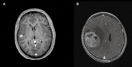
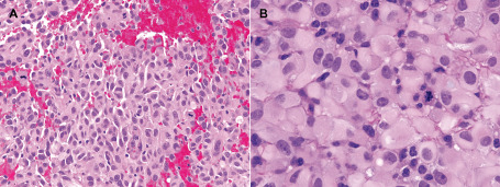
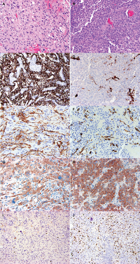
Epithelioid glioblastoma (eGBM) and pleomorphic xanthoastrocytoma (PXA) with anaplastically transformed foci (ePXA) show overlapping features. Eleven eGBMs and 5 ePXAs were reviewed and studied immunohistochemically. Fluorescence in situ hybridization for EGFR amplification, PTEN deletion and ODZ3 deletion was also performed, with Ilumina 450 methylome analysis obtained in five cases. The average age for eGBM was 30.9 (range 2-79) years, including five pediatric cases and a M : F ratio of 4.5. The ePXA patients had a M : F ratio of 4 and averaged 21.2 (range 10-38) years in age, including two pediatric cases. Six eGBMs and two ePXAs recurred (median recurrence interval of 12 and 3.3 months, respectively). All tumors were composed of solid sheets of loosely cohesive, "melanoma-like" cells with only limited infiltration. ePXAs showed lower grade foci with classic features of PXA. Both tumor types showed focal expression of epithelial and glial markers, retained INI1 and BRG1 expression, occasional CD34 positivity, and lack of mutant IDH1 (R132H) immunoreactivity. BRAF V600E mutation was present in four eGBMs and four ePXAs. ODZ3 deletion was detected in seven eGBMs and two ePXAs. EGFR amplification was absent. Methylome analysis showed that one ePXA and one eGBM clustered with PXAs, one eGBM clustered with low-grade gliomas, and two eGBMs clustered with pediatric-type glioblastomas. Common histologic, immunohistochemical, molecular and clinical features found in eGBM and ePXA suggest that they are closely related or the same entity. If the latter is true, the nomenclature and WHO grading remains to be resolved.
Keywords: BRAFV600E; ODZ3; epithelioid; glioblastoma; pediatric; pleomorphic; prognosis; xanthoastrocytoma.
© 2015 International Society of Neuropathology.
Figures

References
-
- Babu R, Hatef J, McLendon RE, Cummings TJ, Sampson JH, Friedman AH, Adamson C (2013) Clinicopathological characteristics and treatment of rhabdoid glioblastoma. J Neurosurg 119:412–419. - PubMed
-
- Chakrabarty A, Mitchell P, Bridges LR, Franks AJ (1999) Malignant transformation in pleomorphic xanthoastrocytoma—a report of two cases. Br J Neurosurg 13:516–519. - PubMed
-
- Chamberlain MC (2013) Salvage therapy with BRAF inhibitors for recurrent pleomorphic xanthoastrocytoma: a retrospective case series. J Neurooncol 114:237–240. - PubMed
MeSH terms
Substances
LinkOut - more resources
Full Text Sources
Medical
Research Materials
Miscellaneous
