Comparative Study
. 2015 Oct 1;75(19):4235-43.
doi: 10.1158/0008-5472.CAN-15-0964. Epub 2015 Aug 3.

Colon Cancer Growth and Dissemination Relies upon Thrombin, Stromal PAR-1, and Fibrinogen

Affiliations
Comparative Study

Colon Cancer Growth and Dissemination Relies upon Thrombin, Stromal PAR-1, and Fibrinogen

Gregory N Adams et al. Cancer Res. .

Abstract

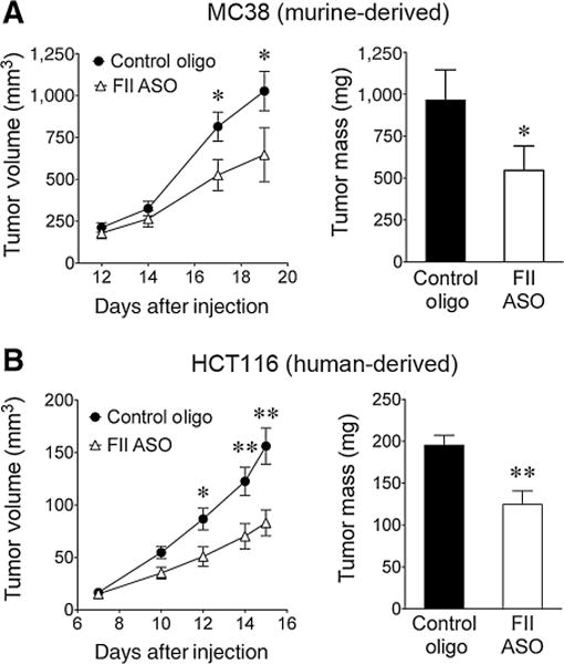
Thrombin-mediated proteolysis is a major determinant of metastasis, but is not universally important for primary tumor growth. Here, we report that colorectal adenocarcinoma represents one important exception whereby thrombin-mediated functions support both primary tumor growth and metastasis. In contrast with studies of multiple nongastrointestinal cancers, we found that the growth of primary tumors formed by murine and human colon cancer cells was reduced in mice by genetic or pharmacologic reduction of circulating prothrombin. Reduced prothrombin expression was associated with lower mitotic indices and invasion of surrounding tissue. Mechanistic investigations revealed that thrombin-driven colonic adenocarcinoma growth relied upon at least two targets of thrombin-mediated proteolysis, protease-activated receptor-1 (PAR-1) expressed by stromal cells and the extracellular matrix protein, fibrinogen. Colonic adenocarcinoma growth was reduced in PAR-1-deficient mice, implicating stromal cell-associated PAR-1 as one thrombin target important for tumor outgrowth. Furthermore, tumor growth was dramatically impeded in fibrinogen-deficient mice, offering the first direct evidence of a critical functional role for fibrinogen in malignant tumor growth. Tumors harvested from fibrinogen-deficient mice displayed a relative reduction in cell proliferative indices, as well as increased tumor necrosis and decreased tumor vascular density. Collectively, our findings established a functional role for thrombin and its targets PAR-1 and fibrinogen in the pathogenesis of colonic adenocarcinoma, supporting tumor growth as well as local invasion and metastasis.


Conflict of interest statement

Disclosure of Potential Conflicts of Interest

B.P. Monia is Senior Vice President and has ownership interest (including patents) in Isis Pharmaceuticals. No potential conflicts of interest were disclosed by the other authors.

Figures

Figure 1
Figure 1
Prothrombin promotes the growth and local invasion of colonic adenocarcinoma in mice. A, tumor growth in WT and FIILox/− mice (n = 5/genotype) following injection of MC38 cells into the dorsal subcutis based on serial caliper measurements and tumor mass at the time of sacrifice (*, P < 0.05; **, P < 0.01). B, shown are representative H&E-stained sections of MC38 tumors harvested from WT and FIILox/− revealing sheets of tumor cells and focal areas of necrosis (dotted lines) in both genotypes. C, analyses at higher magnification revealed that mitotic figures (insets) were more common in tumors harvested from WT mice. This was confirmed by quantitation of mitotic indices (see text for details). D, fibrin(ogen) deposits in tumor tissue (brown immunostaining) were indistinguishable between genotypes. Fibrin(ogen) deposits were generally peritumoral adjacent to overlying dermal tissue, with occasional sparse deposits associated with small areas of necrosis (not shown). E, tumor vascular density was similar between genotypes based on ImageJ analyses of frozen tissue sections immunofluorescently stained for PECAM/CD31. The data shown represents the results of analyses of 6 individual images per tumor from 10 tumors harvested from WT mice and 8 tumors harvested from FIILox/− mice from two experiments with similar outcomes. F, detailed analyses of the tumor/skin junction revealed that local invasion of panniculus muscle fibers (*) was more common in tumors harvested from WT mice relative to those from FIILox/− mice. Also shown are quantitative analyses revealing that local invasion of the panniculus muscle was more common in tumors harvested from WT mice relative to those from FIILox/− mice. This was true even when the analyses were limited to tumors from each genotype of comparable size (~450 mm3). (P < 0.005 for each comparison, Fisher exact test. See text for details.) G, in contrast with observations with colon cancer cells, tumor growth in WT and FIILox/− mice (n = 7/genotype) was indistinguishable following injection of B16 melanoma cells into the dorsal subcutis. Size bars, 100 μm (B, D–F) or 50 μm (C).
Figure 2
Figure 2
Prothrombin is a major determinant of the metastatic potential of colon cancer cells. Cohorts of WT and FIILox/− mice were i.v. injected in parallel via the tail vein with 5 × 10⁵ GFP-expressing MC38 cells. A, shown are representative high-power views of micrometastatic pulmonary foci visualized using a fluorescence-equipped stereomicroscope from WT and FIILox/− mice 30 minutes after injection. Quantitative analyses of micrometastases from 10 high-powered fields per lung demonstrated similarly abundant foci in both genotypes at this early time point. B, gross appearance of representative lung lobes harvested 20 hours after tumor cell injection. Here, quantitation of total surface pulmonary micrometastases revealed significantly fewer foci in lungs harvested from mice with low prothrombin expression relative to control animals. C, analyses 2 weeks after injection demonstrated multiple macroscopic pulmonary foci in the majority of lungs harvested from WT mice, whereas 8 of 10 lungs harvested from FIILox/− mice had no discernable metastatic foci whatsoever. Horizontal bars represent medians; size bars, 250 μm (A) or 2 mm (B and C).
Figure 3
Figure 3
Pharmacologic inhibition of hepatic prothrombin production impedes murine and human colon cancer growth. A, diminution of prothrombin levels to approximately 5% of normal with a prothrombin-specific ASO (n = 10) significantly impeded the growth of MC38 cells in C57Bl/6 mice relative to administration of a control oligonucleotide with no homology in the murine genome (n = 13) as determined by serial caliper measurements estimating tumor volume and tumor mass determined at sacrifice. B, diminishing prothrombin expression in male athymic nude mice (n = 13) also significantly impeded the growth of the human colon cancer cell line HCT116 relative to administration of a control oligonucleotide (n = 9; *, P < 0.05; **, P < 0.01).
Figure 4
Figure 4
Stromal cell–associated PAR-1 promotes colon cancer growth. A, comparison of tumor growth following injection of MC38 cells into the dorsal subcutis of WT and PAR1−/− mice; n = 8/cohort; *, P < 0.05. B, qualitative tumor vasculature as visualized by immunofluorescence staining using an anti-PECAM antibody as well as quantitative measurements of vascular density based on ImageJ analyses of 10 images per tumor were similar between genotypes. C, qualitative and quantitative analyses of TAMs based on immunofluorescent staining of CD68 were also similar in tumors harvested from WT and PAR1−/− mice. Note that TAMs were quantitated as a function of the total tumor tissue area from 8 images taken from each tumor using ImageJ software; size bars, 100 μm.
Figure 5
Figure 5
Fibrinogen promotes tumor progression. A, tumor growth was significantly impeded following injection of MC38 cells into the dorsal subcutis of female Fib mice (n = 6) relative to Fib+ mice (n = 10). B, shown are representative low- and high-power images of H&E-stained tumor sections harvested from Fib+ and Fib mice 20 days after inoculation. Note that areas of necrosis (dotted line in low power images) were more common in tumors harvested from Fib mice and that mitotic figures (insets in high power images) were more prevalent in tumors harvested from Fib+ mice. C, quantitation of necrotic tumor tissue as a function of tumor tissue surface area analyzed (see Materials and Methods) and mitotic indices confirmed that colonic adenocarcinomas harvested from Fib mice were more necrotic and less proliferative than tumors from control animals. D, shown are representative sections of tumor tissue harvested 20 days after tumor cell injection immunofluorescently stained for PECAM as well as the adjacent H&E-stained section revealing healthy appearing tumor tissue. E, tumor vascular density and mean vessel size assessed by quantitation of PECAM-staining area from 7 images per tumor using ImageJ software were significantly diminished in tumors harvested from Fib relative to those harvested from Fib+ mice. F, results of a separate experiment showing the mass of MC38 tumors harvested from female Fib+ (n = 8) and Fib mice (n = 7) 14 days after inoculation. Shown are representative sections of tumor tissue harvested at this earlier time point immunofluorescently stained for PECAM. G, in contrast with analyses performed after a longer period of tumor growth, tumor vasculature density and vessel size were similar in tumors harvested from Fib+ and Fib mice at this early time point; *, P < 0.05; **, P < 0.01; §, P < 0.001; size bars, 200 μm (B low power, D and F) or 20 μm (B high power).

References

    1. Palumbo JS. Mechanisms linking tumor cell-associated procoagulant function to tumor dissemination. Semin Thromb Hemost. 2008;34:154–60. - PubMed
    1. Palumbo JS, Talmage KE, Massari JV, La Jeunesse CM, Flick MJ, Kombrinck KW, et al. Tumor cell-associated tissue factor and circulating hemostatic factors cooperate to increase metastatic potential through natural killer cell-dependent and -independent mechanisms. Blood. 2007;19:19. - PMC - PubMed
    1. Ruf W. Tissue factor and cancer. Thromb Res. 2012;130(Suppl 1):S84–7. - PMC - PubMed
    1. Kasthuri RS, Taubman MB, Mackman N. Role of tissue factor in cancer. J Clin Oncol. 2009;27:4834–8. - PMC - PubMed
    1. Horowitz NA, Blevins EA, Miller WM, Perry AR, Talmage KE, Mullins ES, et al. Thrombomodulin is a determinant of metastasis through a mechanism linked to the thrombin binding domain but not the lectin-like domain. Blood. 2011;118:2889–95. - PMC - PubMed

Publication types

MeSH terms