Review
. 2015 Aug 3;5(8):a021378.
doi: 10.1101/cshperspect.a021378.

Hepatitis B Virus and Hepatitis D Virus Entry, Species Specificity, and Tissue Tropism

Affiliations
Review

Hepatitis B Virus and Hepatitis D Virus Entry, Species Specificity, and Tissue Tropism

Koichi Watashi et al. Cold Spring Harb Perspect Med. .

Abstract

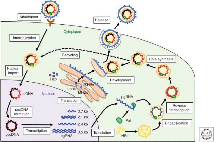
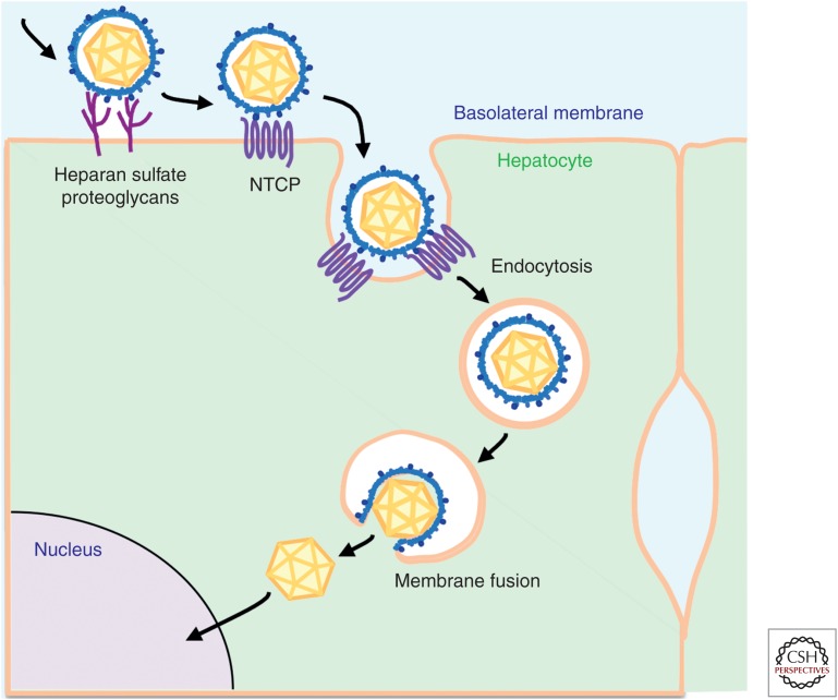
Entry of hepatitis B (HBV) and hepatitis D viruses (HDV) into a host cell represents the initial step of infection. This process requires multiple steps, including the low-affinity attachment of the virus to the cell surface, followed by high-affinity attachment to specific receptor(s), and subsequent endocytosis-mediated internalization. Within the viral envelope, the preS1 region is involved in receptor binding. Recently, sodium taurocholate cotransporting polypeptide (NTCP) has been identified as an entry receptor of HBV and HDV by affinity purification using a preS1 peptide. NTCP is mainly or exclusively expressed in the liver, and this membrane protein is at least one of the factors determining the narrow species specificity and hepatotropism of HBV and HDV. However, there are likely other factors that mediate the species and tissue tropism of HBV. This review summarizes the current understanding of the mechanisms of HBV/HDV entry.


Figures

Figure 1.
Figure 1.
The life cycle of hepatitis B virus (HBV). HBV attaches to host hepatocytes, and subsequent entry steps follow, including internalization and membrane fusion. After nuclear import, the relaxed circular HBV DNA genome is converted into covalently closed circular DNA (cccDNA). HBV replication proceeds with steps, including transcription, translation, encapsidation, reverse transcription, DNA synthesis, envelopment, release, and recycling. Only certain cell cultures can reproduce the steps from attachment to cccDNA formation, such as primary human (PHH) and tupaia (PTH) hepatocytes, differentiated HepaRG cells, and human hepatocyte-derived cell lines complemented with human sodium taurocholate cotransporting polypeptide (hNTCP). HBc, HBV core proteins; LHBs, large HBV surface proteins; MHBs, medium HBV surface proteins; pgRNA, pregenomic RNA; rcDNA, relaxed circular DNA; SHBs, small HBV surface proteins.
Figure 2.
Figure 2.
Structure of hepatitis B virus (HBV) surface proteins. Large HBV surface proteins (LHBs), medium HBV surface proteins (MHBs), and small HBV surface proteins (SHBs) consist of 389–400, 281, and 226 aa (amino acids), respectively. SHBs have four transmembrane domains (I–IV). LHBs are myristoylated (myr) at its amino-terminal end, which is critically involved in HBV and HDV infection. The aa 2–48 portion of the preS1 region is essential for viral infection through receptor binding. The antigenic loop within SHBs also plays a significant role in viral infection.
Figure 3.
Figure 3.
Schematic representation of the HBV entry pathway. HBV particles attach to host hepatocytes through cell-surface factors, including heparan sulfate proteoglycans (HSPGs) with low affinity. The particles then interact with higher affinity with a specific receptor, sodium taurocholate cotransporting polypeptide (NTCP), to mediate the ensuing internalization. Internalization occurs in an endocytosis-dependent manner. The viral and cellular membranes are fused in certain vesicles and the nucleocapsids inside the viral particles are released into the cytoplasm, and are then destined to traffic to the nucleus for translocation via the nuclear pore complex.

References

    1. Abou-Jaoude G, Sureau C. 2007. Entry of hepatitis delta virus requires the conserved cysteine residues of the hepatitis B virus envelope protein antigenic loop and is blocked by inhibitors of thiol-disulfide exchange. J Virol 81: 13057–13066. - PMC - PubMed
    1. Araki K, Miyazaki J, Hino O, Tomita N, Chisaka O, Matsubara K, Yamamura K. 1989. Expression and replication of hepatitis B virus genome in transgenic mice. Proc Natl Acad Sci 86: 207–211. - PMC - PubMed
    1. Barrera A, Guerra B, Notvall L, Lanford RE. 2005. Mapping of the hepatitis B virus pre-S1 domain involved in receptor recognition. J Virol 79: 9786–9798. - PMC - PubMed
    1. Bremer CM, Sominskaya I, Skrastina D, Pumpens P, El Wahed AA, Beutling U, Frank R, Fritz HJ, Hunsmann G, Gerlich WH, et al. 2011. N-terminal myristoylation-dependent masking of neutralizing epitopes in the preS1 attachment site of hepatitis B virus. J Hepatol 55: 29–37. - PubMed
    1. Bruss V, Hagelstein J, Gerhardt E, Galle PR. 1996. Myristylation of the large surface protein is required for hepatitis B virus in vitro infectivity. Virology 218: 396–399. - PubMed

Publication types