. 2015 Aug 4:6:7939.
doi: 10.1038/ncomms8939.

A paclitaxel-loaded recombinant polypeptide nanoparticle outperforms Abraxane in multiple murine cancer models

Affiliations

A paclitaxel-loaded recombinant polypeptide nanoparticle outperforms Abraxane in multiple murine cancer models

Jayanta Bhattacharyya et al. Nat Commun. .

Abstract

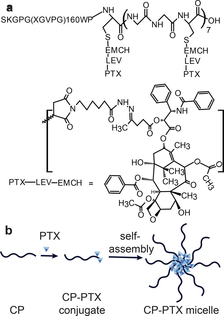
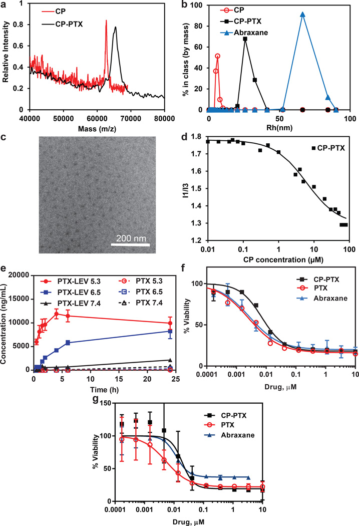
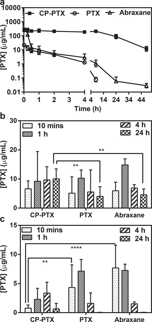
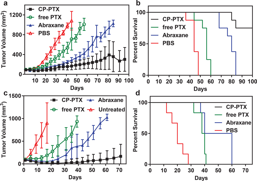
Packaging clinically relevant hydrophobic drugs into a self-assembled nanoparticle can improve their aqueous solubility, plasma half-life, tumour-specific uptake and therapeutic potential. To this end, here we conjugated paclitaxel (PTX) to recombinant chimeric polypeptides (CPs) that spontaneously self-assemble into ∼60 nm near-monodisperse nanoparticles that increased the systemic exposure of PTX by sevenfold compared with free drug and twofold compared with the Food and Drug Administration-approved taxane nanoformulation (Abraxane). The tumour uptake of the CP-PTX nanoparticle was fivefold greater than free drug and twofold greater than Abraxane. In a murine cancer model of human triple-negative breast cancer and prostate cancer, CP-PTX induced near-complete tumour regression after a single dose in both tumour models, whereas at the same dose, no mice treated with Abraxane survived for >80 days (breast) and 60 days (prostate), respectively. These results show that a molecularly engineered nanoparticle with precisely engineered design features outperforms Abraxane, the current gold standard for PTX delivery.


Conflict of interest statement

Conflict of Interest A.C. has a financial interest in a start-up company, PhaseBio Pharmaceuticals that has licensed the technology reported herein from Duke University.

Figures

Figure 1
Figure 1. Structure of CP–PTX conjugate and schematic of the structure of CP-PTX nanoparticles
a, The CP was synthesized by genetically encoded synthesis in E. coli, and conjugated to PTX at the multiple Cys residues at the C-terminal end of the CP by a pH sensitive linker. b, Attachment of the hydrophobic drug PTX triggers self-assembly of the CP into spherical nanoparticles with a drug-rich (blue triangles) core surrounded by a hydrophilic polypeptide corona (black chains).
Figure 2
Figure 2. Characterization of CP–PTX nanoparticles
a. MALDI-MASS of CP and CP-PTX conjugate. b-d, Determination of hydrodynamic radius (b), cryo-TEM (c), and critical aggregation concentration (d) of CP-PTX conjugate. e, The kinetics of pH-dependent release of PTX from CP–PTX nanoparticles as determined by LCMS/MS at pH 7.4, 6.5 and 5.3 (mean ± SD). f-g, Cell viability for CP–PTX and free PTX in MDA-MB-231 (f) and PC3 (g) cells (mean ± 95%CI).
Figure 3
Figure 3. Cell cycle arrest at the G2/M phase by CP-PTX
a, MDA-MB-231 cells were treated with 1 nM PTX equivalent at 37 °C for 48 h. Adherent cells were collected, fixed in 70% ethanol, treated with RNase A and stained with propidium iodide (PI). Samples were analyzed by flow cytometry. Results are expressed as the percentage of each subpopulation as determined by analysis of 5,000 cells per sample. (Two way ANOVA, mean ± 95% CI, n=3, p< 0.05). b, Stabilization of microtubule structure by CP-PTX in MDA-MB-231 cells. Cells were treated with 1 nM CP-PTX or free PTX at 37 °C. After 24 h, cells were stained with α-tubulin antibody (green), Hoechst 33342 (blue) and WGA (red) and imaged by confocal fluorescence microscopy. Scale bar, 10 µm.
Figure 4
Figure 4. Plasma pharmacokinetics and tissue biodistribution
a, Plasma PTX concentration as a function of time post-injection. A non-compartment model was fitted to the plasma PTX concentration, which yielded a terminal half-life of 8.4 h for CP–PTX and 3.8 h for Abraxane (mean ± 95% CI, n=4). b, c, The PTX concentration in tumor (b) and muscle (c) at 10 min and 24 h post-administration of free PTX, Abraxane and CP-PTX nanoparticles. ** and **** indicates p<0.01 and p<0.0001 respectively (Two way ANOVA, Tukey’s test) (mean ± 95% CI, n=4).
Figure 5
Figure 5. Anti-tumor activity of CP–PTX nanoparticles
a, and b, tumor cells (MDA-MB-231) were implanted in the mammary fat pad on day zero. When the tumor volume reached ~100 mm3, mice were treated at the MTD with PBS (n=8), free PTX (25 mg kg−1 BW, n=8) or CP–PTX (50 mg PTX equiv.kg−1 BW, n=8). a, Tumor volume up to day 100 (mean ± 95% CI, n= 8). p = 0.0001 for CP–PTX versus PTX and PBS (day 44) respectively (Tukey test). b, Cumulative survival of mice (Kaplan–Meier). c-d, Tumor cells (PC3) were implanted on the right flank on day zero. When the tumor volume reached 50-100 mm3, mice were treated at the MTD with PBS (n=6), free PTX (25 mg kg−1 BW, n=6) and CP–PTX (50 mg PTX equiv.kg−1BW, n=6). c, Tumor volume up to day 70 (mean= ±95%CI, n=6). p=0.0001 for CP–PTX versus PTX and PBS (day 30) respectively (Tukey’s test). d, Cumulative survival of mice (Kaplan–Meier).
Figure 6
Figure 6. Gene expression analysis of tumors treated with CP-PTX or PTX
a, The global gene expression of xenografts that are responsive to treatment with CP-PTX nanoparticle or PTX or relapse upon treatment, was determined using microarray analysis using U133A2 (Affymetrix) arrays. The transcriptional response of each treatment was derived by zero-transformation. Selected gene clusters that were induced and repressed specifically in the CP-PTX tumors that responded to treatment are highlighted by the color bar. b, The three gene clusters that reflected the distinct changes in the CP-PTX response tumors were further expanded with selected names shown.

References

    1. Scott RB. Cancer chemotherapy-the first twenty-five years. Br. Med. J. 1970;4:259–265. - PMC - PubMed
    1. Zhang J, Yang PL, Gray NS. Targeting cancer with small molecule kinase inhibitors. Nat. Rev. Cancer. 2009;9:28–39. - PubMed
    1. Lee CC, MacKay JA, Frechet JMJ, Szoka FC. Designing dendrimers for biological applications. Nature Biotechnol. 2005;23:1517–1526. - PubMed
    1. Matsumura Y, Maeda H. A new concept for macromolecular therapeutics in cancer chemotherapy: Mechanism of tumoritropic accumulation of proteins and the antitumor agent smancs. Cancer Res. 1986;46:6387–6392. - PubMed
    1. McDaniel JR, et al. Self-Assembly of Thermally Responsive Nanoparticles of a Genetically Encoded Peptide Polymer by Drug Conjugation. Angew. Chem. Int. Ed. 2013;52:1683–1687. - PMC - PubMed

Publication types

MeSH terms