. 2016 Feb;117(2):491-9.
doi: 10.1002/jcb.25300.

Schwann Cells Increase Prostate and Pancreatic Tumor Cell Invasion Using Laminin Binding A6 Integrin

Affiliations

Schwann Cells Increase Prostate and Pancreatic Tumor Cell Invasion Using Laminin Binding A6 Integrin

Isis C Sroka et al. J Cell Biochem. 2016 Feb.

Abstract

Human pancreatic and prostate cancers metastasize along nerve axons during perineural invasion. The extracellular matrix laminin class of proteins is an abundant component of both myelinated and non-myelinated nerves. Analysis of human pancreatic and prostate tissue revealed both perineural and endoneural invasion with Schwann cells surrounded or disrupted by tumor, respectively. Tumor and nerve cell co-culture conditions were used to determine if myelinating or non-myelinating Schwann cell (S16 and S16Y, respectively) phenotype was equally likely to promote integrin-dependent cancer cell invasion and migration on laminin. Conditioned medium from S16 cells increased tumor cell (DU145, PC3, and CFPAC1) invasion into laminin approximately 1.3-2.0 fold compared to fetal bovine serum (FBS) treated cells. Integrin function (e.g., ITGA6p formation) increased up to 1.5 fold in prostate (DU145, PC3, RWPE-1) and pancreatic (CFPAC1) cells, and invasion was dependent on ITGA6p formation and ITGB1 as determined by function-blocking antibodies. In contrast, conditioned medium isolated from S16Y cells (non-myelinating phenotype) decreased constitutive levels of ITGA6p in the tumor cells by 50% compared to untreated cells and decreased ITGA6p formation 3.0 fold compared to S16 treated cells. Flow cytometry and western blot analysis revealed loss of ITGA6p formation as reversible and independent of overall loss of ITGA6 expression. These results suggest that the myelinating phenotype of Schwann cells within the tumor microenvironment increased integrin-dependent tumor invasion on laminin.

Keywords: A6B1; LAMININ; METASTASIS; MICROENVIRONMENT; PERINEURAL INVASION; SCHWANN CELLS.


Conflict of interest statement

Conflict of interest: None.

Figures

Fig. 1
Fig. 1
Perineural and endoneural tumor cell invasion in human prostate and pancreatic cancer. (A) Prostate cancer cells (Ca) invade around the perineurium of the nerve (N) near Schwann cells (SCs, arrow; stained with S100 antibody; magnification × 20). (B) Endoneural invasion shows SCs stained with S100 antibody; interspersed throughout the invaded nerve (arrow) and in contact with prostate tumor cells (× 40). (C) Pancreatic cancer tissue stained with the S100 antibody indicating the presence of SCs in the nerve (arrow) with cancer invading the perineurium (× 20). (D) ITGA6 expression (dark brown) on pancreatic cancer cells (× 20) invading the perineurium of the same nerve depicted in part C and within SCs as expected.
Fig. 2
Fig. 2
Immortalized S16 and S16Y rat Schwann cell lines expressed Schwann cell specific markers. Immunoblot analysis was used to identify expression of the Schwann cell specific transcription factor SOX10 and the S100β protein. Actin was used as a loading control. The results are representative of three independent experiments.
Fig. 3
Fig. 3
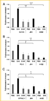
S16 conditioned media increased tumor cell invasion dependent on A6B1. (A) DU145, (B) PC3, and (C) CFPAC1 cells were analyzed using the Cultrex modified Boyden chamber invasion assay with laminin 111. The fold-increase in invasion was determined under conditions of either FBS, S16 (black bars), or S16Y (gray bars) conditioned media, FBS+J8H (ITGA6 blocking antibody), S16+J8H (ITGA6 blocking antibody), FBS+AIIB2 (ITGB1 blocking antibody), or S16+AIIB2 (ITGB1 blocking antibody). The cells were treated with the blocking antibodies during the invasion assay. The results are expressed as mean values ± SD of three independent experiments. The asterisks denote a significant difference (*P < 0.05; **P < 0 .005; ***P < 0 .0001, unpaired Student Test) comparing the samples as indicated by the brackets.
Fig. 4
Fig. 4
Suppression of ITGA6p production in tumor cells by S16Y cell (non-myelinating phenotype) conditioned media. (A) DU145, PC3, CFPAC1 tumor, and normal prostate (RWPE-1) cells were treated with DMEM control media (C), or S16 and S16Y conditioned media for 24 h. Integrin A6 (ITGA6) and A6p (ITGA6p) were immunoprecipitated using the J1B5 antibody and detected by immuno blot. (B) Quantitative analysis of the immuno blot experiments in part A for DU145, PC3, CFPAC1, and RWPE-1 cell lines. NIH Image J analysis determined the ratio of area density of ITGA6p to ITGA6 as shown. The results are representative of three independent experiments and the asterisk denotes a significant difference (P < 0.05, unpaired Student T-test) as compared to the control media (C) sample. (C) RWPE and DU145 cells were treated with DMEM control media (C) or 100 uM amiloride (Am) or S16 conditioned media alone (S16) or S16 conditioned media containing amiloride (S16 Am) for 24 h. Immunoblot analysis of whole cell lysates was used to detect integrin A6 (ITGA6) or the ITGA6-NT fragment as a surrogate marker for ITGA6p production. NIH Image J analysis determined the ratio of area density of ITGA6p to ITGA6 as shown. The results are representative of three independent experiments and the asterisk denotes a significant difference (P < 0.05, unpaired Student T-test) as compared to the signal in the control sample of each group.
Fig. 5
Fig. 5
Recovery of ITGA6p expression in tumor cell lines treated with S16Y (non-myelinating phenotype) conditioned medium. (A) The DU145, the (B) PC3, and (C) CFPAC1 cancer cell lines were treated with S16Y conditioned medium for 24 h to decrease ITGA6p expression. The ITGA6NT fragment is a surrogate marker for ITGA6p production. The cells were then treated with either S16Y medium (S16Y M) or control medium (IMDM) for an additional 24, 48, or 72 h. Immunoblot analysis of whole cell lysates was used to detect A6 (ITGA6) and ITGA6NT with actin used as a loading control (A, B, and C, panel a). Quantitative analysis of the immunoblots A, B, and C is shown in (b). NIH Image J analysis determined the ratio of area density of ITGA6p to ITGA6 as shown. The results are representative of three independent experiments and the asterisk denotes a significant difference (P < 0.05, unpaired Student T-test) as compared to the signal in the 24 h sample of each group. Panel c of A, B, and C is total cell surface levels of ITGA6 as detected by flow analysis on the cell lines treated with control media, S16, or S16Y conditioned media for 24 h. The black peak = cells only control, red peak = cells treated with control media, blue = cells treated with S16 media, and green = cells treated S16Y conditioned media. All results are representative of three independent experiments.

References

    1. Bapat AA, Hostetter G, Von Hoff DD, Han H. Perineural invasion and associated pain in pancreatic cancer. Nat Rev Cancer. 2011;11(10):695–707. - PubMed
    1. Bohnsack JF, Akiyama SK, Damsky CH, Knape WA, Zimmerman GA. Human neutrophil adherence to laminin in vitro. Evidence for a distinct neutrophil integrin receptor for laminin. J Exp Med. 1990;171(4):1221–1237. - PMC - PubMed
    1. Campana WM. Schwann cells: Activated peripheral glia and their role in neuropathic pain. Brain, behavior, and immunity. 2007;21(5):522–527. - PMC - PubMed
    1. Ceyhan GO, Demir IE, Altintas B, Rauch U, Thiel G, Muller MW, Giese NA, Friess H, Schafer KH. Neural invasion in pancreatic cancer: A mutual tropism between neurons and cancer cells. Biochemi Biophys Res Commun. 2008;374(3):442–447. - PubMed
    1. Chernousov MA, Yu WM, Chen ZL, Carey DJ, Strickland S. Regulation of Schwann cell function by the extracellular matrix. Glia. 2008;56(14):1498–1507. - PubMed

Publication types

LinkOut - more resources