. 2015 Sep 15;128(18):3435-43.
doi: 10.1242/jcs.172031. Epub 2015 Aug 3.

Vinculin phosphorylation at residues Y100 and Y1065 is required for cellular force transmission

Affiliations

Vinculin phosphorylation at residues Y100 and Y1065 is required for cellular force transmission

Vera Auernheimer et al. J Cell Sci. .

Abstract

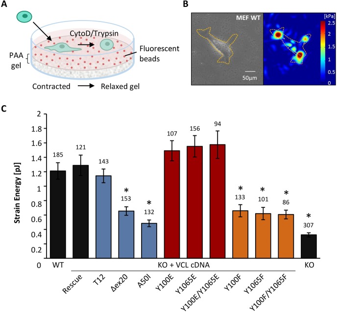
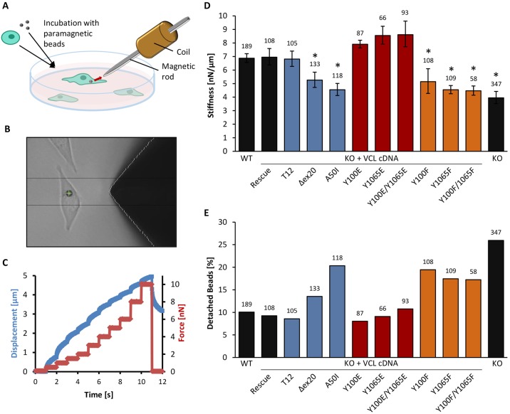
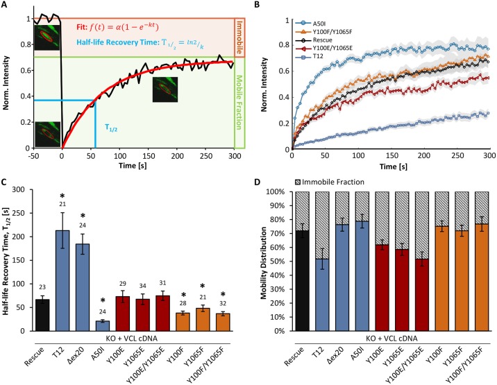
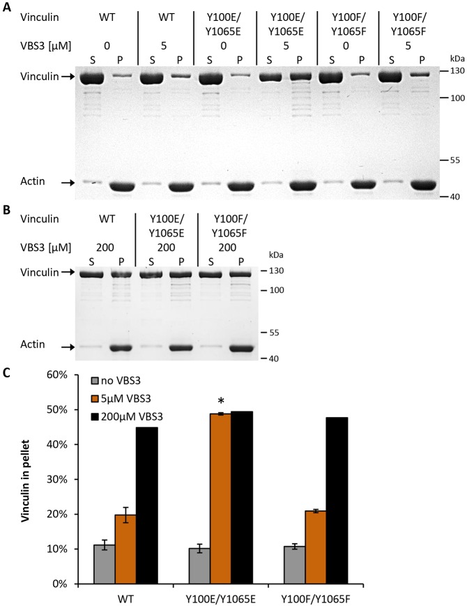
The focal adhesion protein vinculin connects the actin cytoskeleton, through talin and integrins, with the extracellular matrix. Vinculin consists of a globular head and tail domain, which undergo conformational changes from a closed auto-inhibited conformation in the cytoplasm to an open conformation in focal adhesions. Src-mediated phosphorylation has been suggested to regulate this conformational switch. To explore the role of phosphorylation in vinculin activation, we used knock-out mouse embryonic fibroblasts re-expressing different vinculin mutants in traction microscopy, magnetic tweezer microrheology, FRAP and actin-binding assays. Compared to cells expressing wild-type or constitutively active vinculin, we found reduced tractions, cytoskeletal stiffness, adhesion strength, and increased vinculin dynamics in cells expressing constitutively inactive vinculin or vinculin where Src-mediated phosphorylation was blocked by replacing tyrosine at position 100 and/or 1065 with a non-phosphorylatable phenylalanine residue. Replacing tyrosine residues with phospho-mimicking glutamic acid residues restored cellular tractions, stiffness and adhesion strength, as well as vinculin dynamics, and facilitated vinculin-actin binding. These data demonstrate that Src-mediated phosphorylation is necessary for vinculin activation, and that phosphorylation controls cytoskeletal mechanics by regulating force transmission between the actin cytoskeleton and focal adhesion proteins.

Keywords: Actin pulldown; Cell stiffness; FRAP; Focal adhesion; Mechanotransduction; Traction; Tyrosine phosphorylation; Vinculin.


Conflict of interest statement

Competing interests

The authors declare no competing or financial interests.

Figures

Fig. 1.
Fig. 1.
Measurements of contractile forces by 2D traction microscopy. (A) Schematic representation of the 2D traction setup. Cells attach to fibronectin-coated PAA gels with embedded fluorescent beads. The cells are detached by using cytochalasin D and trypsin, and the elastic gel relaxes to its original position. The bead positions are recorded before and after the relaxation, and tractions forces are calculated from the displacement. (B) A bright-field image (left) and corresponding force map (right) of a wild-type (WT) MEF cell on a PAA gel. The dashed lines outline the cell. The color bar indicates tractions in kPa. Scale bar: 50 µm. (C) Strain energy values are shown for cells expressing vinculin conformational mutants (blue), vinculin phospho-mimicking (red), non-phosphorylatable mutants (orange), and vinculin WT, rescue and KO cells (black). Contractile forces are reduced for impaired actin-binding capacity (Δex20), the inactive conformation (A50I) and after inhibition of phosphorylation (Y100F, Y1065F, Y100F/Y1065F) of the vinculin molecule. Results are means±s.e.m.; the number of analyzed cells is shown above each column. *P≤0.05 compared to rescue cells (Mann–Whitney U-test).
Fig. 2.
Fig. 2.
Cell stiffness and adhesive forces determined by magnetic tweezer experiments. (A) Schematic representation of the magnetic tweezer setup. Cells are seeded overnight on a cell culture dish and incubated with ECM-coated paramagnetic beads 30 min prior to measurements. A magnetic field is applied to the bead attached to integrins, and the displacement of the bead is recorded. (B) Bright-field image of the magnetic tweezer tip close to a tracked magnetic bead (green cross) attached to the cell. (C) Increasing bead displacement (blue) with increasing force steps at 1 nN/s (red). (D) Stiffness values at 6 nN force are shown for cells expressing vinculin conformational (blue), phospho-mimicking (red) and non-phosphorylatable mutants (orange), as well as for vinculin wild-type (WT), rescue and KO cells (black). The cell stiffness decreases significantly for actin-binding deficient (Δex20), inactive (A50I), non-phosphorylatable (Y100F, Y1065F, Y100F/Y1065F) vinculin mutants and KO cells. Phospho-mimicking mutants (Y100E, Y1065E, Y100E/Y1065E) show slightly higher values than WT, rescue and T12 cells. (E) The percentage of detached beads at 6 nN force was increased for the vinculin mutants Δex20, A50I, Y100F, Y1065F, Y100F/Y1065F and KO cells compared to vinculin WT, rescue, T12 and phospho-mimicking mutants. Results are means±s.e.m.; the number of analyzed cells is shown above each column. *P≤0.05 compared to rescue cells (Mann–Whitney U-test).
Fig. 3.
Fig. 3.
Fluorescence recovery after photobleaching (FRAP) of different vinculin mutants. (A) Recovery curve of a bleached vinculin rescue cell. Intensity of the bleached area is plotted over time, and the single exponential fit is shown in red. Images of the bleached focal adhesion are shown at t=−4 s, t=0 s and t=160 s. (B) Fluorescence recovery of different vinculin mutants (rescue, T12, A50I, Y100E/Y1065E, Y100F/Y1065F) after photobleaching (mean, with the s.e.m. shown by the region shaded gray; the number of analyzed cells is shown above each column in C). (C) The half-life recovery time, T1/2 is significantly increased for the active, open conformations T12 and Δex20 and decreased for the inactive, closed (A50I), and non-phosphorylatable vinculin (Y100F, Y1065F, Y100F/Y1065F) mutants and KO cells compared to rescue cells and phospho-mimicking (Y100E, Y1065E, Y100E/Y1065E) mutants. (D) The mobility distribution shows the percentage of mobile and immobile vinculin proteins in focal adhesions. The immobile fraction is increased for T12 and phospho-mimicking mutants (Y100E, Y1065E, Y100E/Y1065E) compared to rescue cells and Δex20, A50I, Y100F, Y1065F and Y100F/Y1065F mutants. Results are means±s.e.m.; the number of analyzed cells is shown above each column in C. *P≤0.05 compared to rescue cells (Mann–Whitney U-test).
Fig. 4.
Fig. 4.
Co-sedimentation assays of vinculin mutants with F-actin in the presence or absence of VBS3. (A) Purified wild-type (WT) vinculin and the indicated vinculin mutants were incubated with F-actin in the presence or absence of 5 µM VBS3. After ultracentrifugation, the supernatant (S) and pellet (P) fractions were separated, and the amount of proteins was analyzed by SDS-PAGE. The amount of vinculin in the pellet fraction only increased for the Y100E/Y1065E mutant. (B) Co-sedimentation of vinculin with F-actin in the presence of 200 µM VBS3. (C) The graph shows the percentage of vinculin in the pellet for all mutants under three different conditions (0 µM VBS3 in gray, 5 µM VBS3 in orange and 200 µM VBS3 in black) after quantitative analysis of the protein bands (n=3). Without VBS3, ∼10% of vinculin co-sedimented with F-actin. Using 5 µM VBS3, the co-sedimentation of WT and Y100F/Y1065F mutant with F-actin increased to 20% and for Y100E/Y1065E mutant up to 50%. Using 200 µM VBS3, all vinculin proteins co-sedimented with F-actin to similar levels (∼50%). Results are means±s.e.m. *P≤0.05 compared to rescue cells (Mann–Whitney U-test).

Similar articles

Cited by

References

    1. Alenghat F. J., Fabry B., Tsai K. Y., Goldmann W. H. and Ingber D. E. (2000). Analysis of cell mechanics in single vinculin-deficient cells using a magnetic tweezer. Biochem. Biophys. Res. Commun. 277, 93-99. 10.1006/bbrc.2000.3636 - DOI - PubMed
    1. Auernheimer V. and Goldmann W. H. (2014). Serine phosphorylation on position 1033 of vinculin impacts cellular mechanics. Biochem. Biophys. Res. Commun. 450, 1095-1098. 10.1016/j.bbrc.2014.06.122 - DOI - PubMed
    1. Bakolitsa C., Cohen D. M., Bankston L. A., Bobkov A. A., Cadwell G. W., Jennings L., Critchley D. R., Craig S. W. and Liddington R. C. (2004). Structural basis for vinculin activation at sites of cell adhesion. Nature 430, 583-586. 10.1038/nature02610 - DOI - PubMed
    1. Bass M. D., Patel B., Barsukov I. G., Fillingham I. J., Mason R., Smith B. J., Bagshaw C. R. and Critchley D. R. (2002). Further characterization of the interaction between the cytoskeletal proteins talin and vinculin. Biochem. J. 362, 761-768. 10.1042/0264-6021:3620761 - DOI - PMC - PubMed
    1. Bays J. L., Peng X., Tolbert C. E., Guilluy C., Angell A. E., Pan Y., Superfine R., Burridge K. and DeMali K. A. (2014). Vinculin phosphorylation differentially regulates mechanotransduction at cell-cell and cell-matrix adhesions. J. Cell Biol. 205, 251-263. 10.1083/jcb.201309092 - DOI - PMC - PubMed

Publication types