. 2015 Sep 15;112(37):11547-52.
doi: 10.1073/pnas.1500964112. Epub 2015 Aug 3.

Oxidative cyclizations in orthosomycin biosynthesis expand the known chemistry of an oxygenase superfamily

Affiliations

Oxidative cyclizations in orthosomycin biosynthesis expand the known chemistry of an oxygenase superfamily

Kathryn M McCulloch et al. Proc Natl Acad Sci U S A. .

Abstract

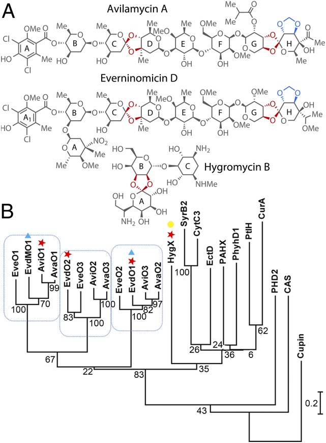
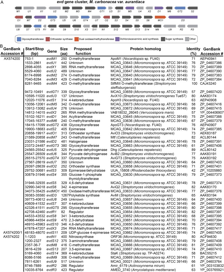
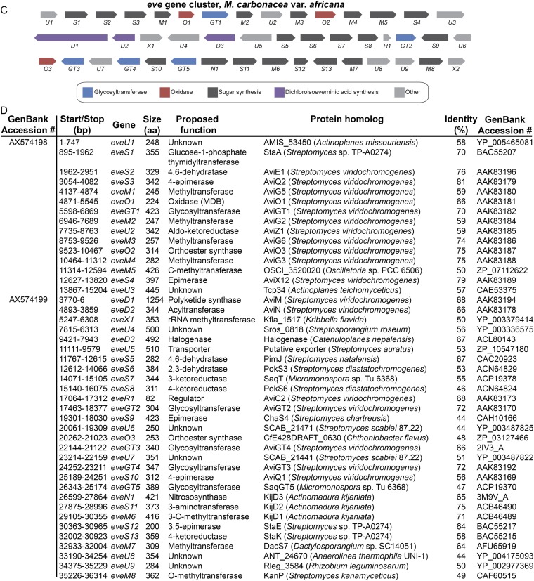
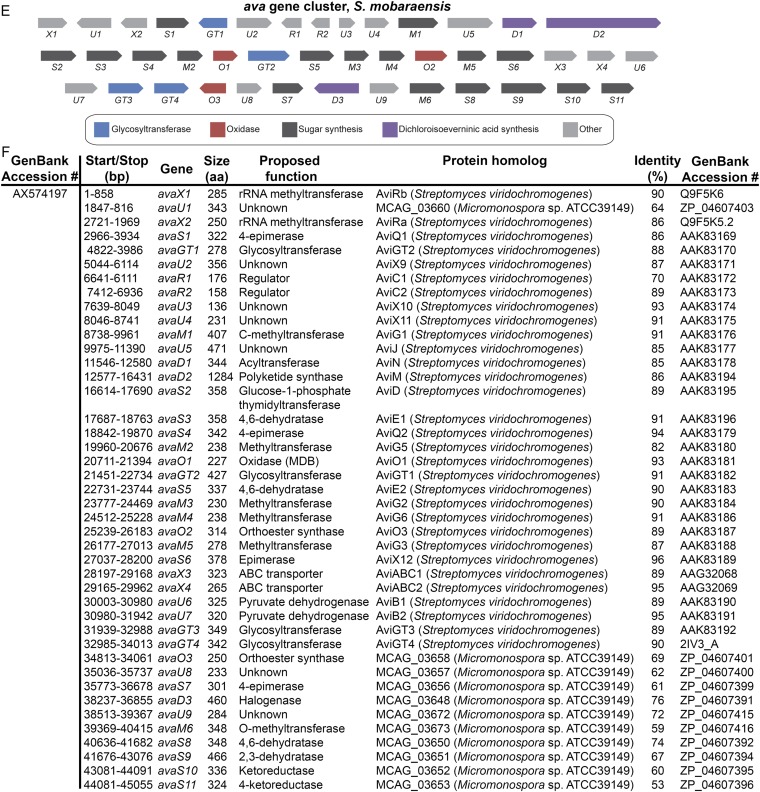
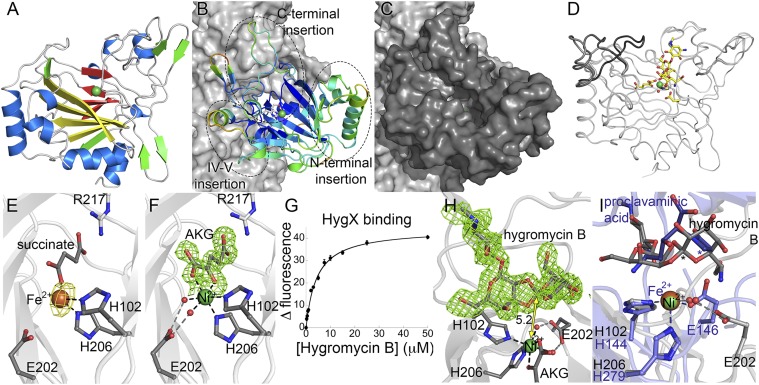
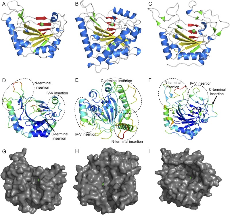
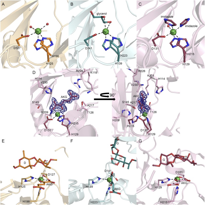
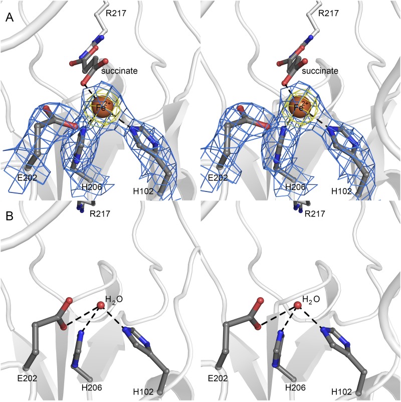
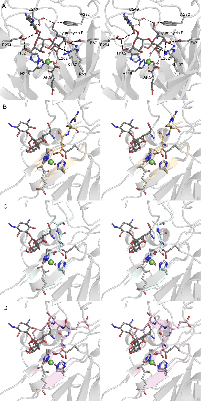
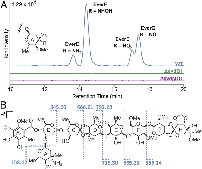
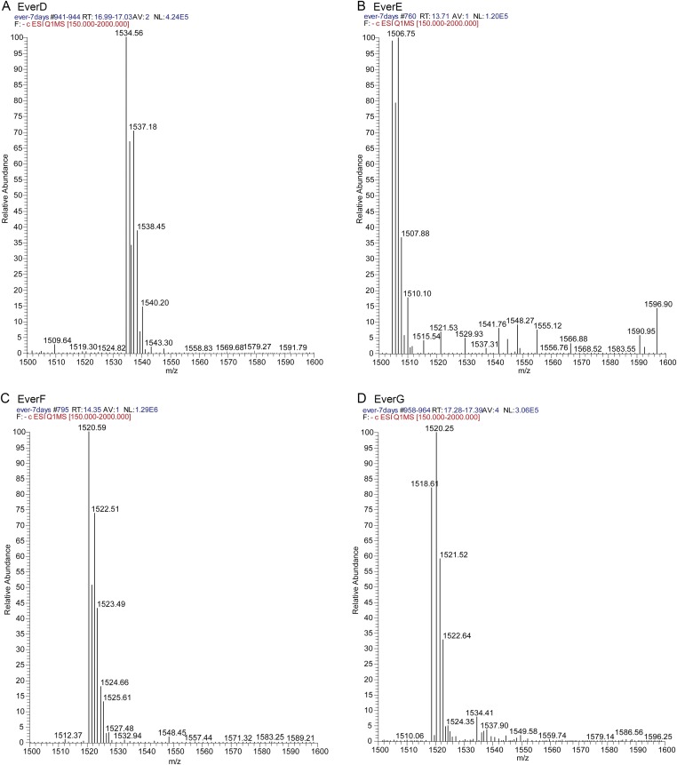
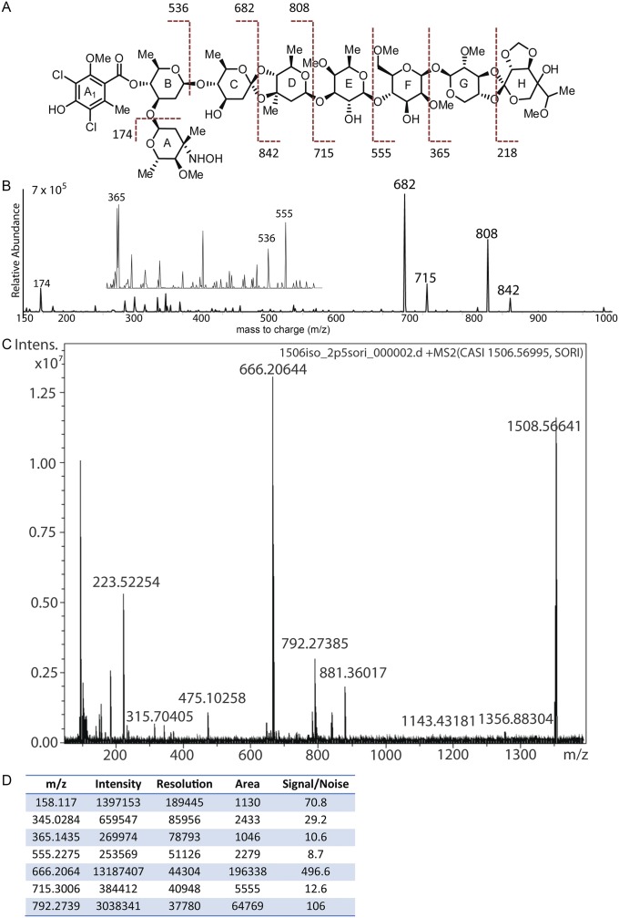
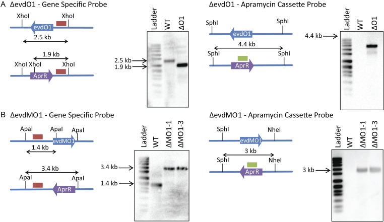
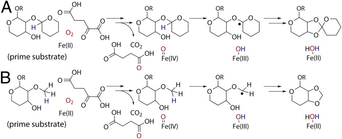
Orthosomycins are oligosaccharide antibiotics that include avilamycin, everninomicin, and hygromycin B and are hallmarked by a rigidifying interglycosidic spirocyclic ortho-δ-lactone (orthoester) linkage between at least one pair of carbohydrates. A subset of orthosomycins additionally contain a carbohydrate capped by a methylenedioxy bridge. The orthoester linkage is necessary for antibiotic activity but rarely observed in natural products. Orthoester linkage and methylenedioxy bridge biosynthesis require similar oxidative cyclizations adjacent to a sugar ring. We have identified a conserved group of nonheme iron, α-ketoglutarate-dependent oxygenases likely responsible for this chemistry. High-resolution crystal structures of the EvdO1 and EvdO2 oxygenases of everninomicin biosynthesis, the AviO1 oxygenase of avilamycin biosynthesis, and HygX of hygromycin B biosynthesis show how these enzymes accommodate large substrates, a challenge that requires a variation in metal coordination in HygX. Excitingly, the ternary complex of HygX with cosubstrate α-ketoglutarate and putative product hygromycin B identified an orientation of one glycosidic linkage of hygromycin B consistent with metal-catalyzed hydrogen atom abstraction from substrate. These structural results are complemented by gene disruption of the oxygenases evdO1 and evdMO1 from the everninomicin biosynthetic cluster, which demonstrate that functional oxygenase activity is critical for antibiotic production. Our data therefore support a role for these enzymes in the production of key features of the orthosomycin antibiotics.

Keywords: antibiotic biosynthesis; crystal structure; nonheme iron α-ketoglutarate–dependent oxygenases; oxidative cyclization.


Conflict of interest statement

The authors declare no conflict of interest.

Figures

Fig. 1.
Fig. 1.
Orthosomycins and associated oxygenases. (A) Orthosomycins with orthoester linkages and methylenedioxy bridges in red. (B) Oxygenase phylogenetic analysis. Enzymes characterized structurally (red ★), through gene disruption (blue ▲), and for binding affinity (yellow ●) are indicated.
Fig. S1.
Fig. S1.
Pictorial annotation and deduced functional assignment for ORFs of the evd, eve, and ava gene clusters. Using antiSMASH (antismash.secondarymetabolites.org), ORFs were identified from GenBank nucleotide sequences. Each ORF was analyzed using Translated BLAST (Blastx). Based on the function of homologous proteins, gene names were assigned and functions of genes were proposed. (A) Pictorial annotation of the evd gene cluster colored by proposed function in orthosomycin biosynthesis. (B) Deduced evd gene functions and nearest homologs for each ORF. (C) Pictorial annotation of the eve gene cluster colored by proposed function in orthosomycin biosynthesis. (D) Deduced eve gene functions and nearest homologs for each ORF. (E) Pictorial annotation of the ava gene cluster colored by proposed function in orthosomycin biosynthesis. (F) Deduced ava gene functions and nearest homologs for each ORF.
Fig. S1.
Fig. S1.
Pictorial annotation and deduced functional assignment for ORFs of the evd, eve, and ava gene clusters. Using antiSMASH (antismash.secondarymetabolites.org), ORFs were identified from GenBank nucleotide sequences. Each ORF was analyzed using Translated BLAST (Blastx). Based on the function of homologous proteins, gene names were assigned and functions of genes were proposed. (A) Pictorial annotation of the evd gene cluster colored by proposed function in orthosomycin biosynthesis. (B) Deduced evd gene functions and nearest homologs for each ORF. (C) Pictorial annotation of the eve gene cluster colored by proposed function in orthosomycin biosynthesis. (D) Deduced eve gene functions and nearest homologs for each ORF. (E) Pictorial annotation of the ava gene cluster colored by proposed function in orthosomycin biosynthesis. (F) Deduced ava gene functions and nearest homologs for each ORF.
Fig. 2.
Fig. 2.
HygX structure. (A) HygX with major strands in yellow and minor strands in red. Ni2+and Fe2+ are green and orange spheres. (B) HygX A chain colored by crystallographic temperature factors where cool colors indicate low B-factors. Loop insertions are highlighted. (C) Surface representation in the same orientation as B. (D) Superposition of four chains of the HygX-AKG complex showing loop movement, with hygromycin B shown for reference. (E) Iron coordination with anomalous difference density in yellow and contoured at 5σ. (F) Active site metal coordination of HygX. |FO| − |FC| difference density is contoured around AKG at 2.5 σ, calculated before inclusion of AKG in the model. (G) Tryptophan fluorescence at 350 nm of 0.5 μM HygX, 0.05 mM NiCl2, 0.05 mM AKG, and varying concentrations of hygromycin B (Kd, 3.4 ± 0.5 μM). (H) 3σ |FO| − |FC| difference density calculated before hygromycin B placement. One possible site of hydrogen atom abstraction is 5.2 Å from the metal center. (I) HygX–AKG–hygromycin B product complex (in gray) aligned with the clavaminate synthase–AKG–proclavaminate substrate complex (blue; PDB ID code 1DRT). Asterisks mark sites of hydrogen atom abstraction.
Fig. S2.
Fig. S2.
Orthosomycin-associated oxygenase structures. Cartoon representations of AviO1 (A), EvdO1 (B), and EvdO2 (C) in the same orientations. The major sheet is colored yellow, the minor sheet is red, and nickel ions are shown as green spheres. (DF) Cartoon representation of AviO1 (D), EvdO1 (E), and EvdO2 (F) colored by crystallographic temperature factors. Warm colors indicate high B-factors and more thermal motion. Three insertion sites common to the PhyH subfamily of AKG/Fe(II)-dependent oxidases are labeled. The orientation of each maximizes the view of the three insertions and the binding site. (GI) Surface representation of AviO1 (G), EvdO1 (H), and EvdO2 (I) showing the prime substrate binding site in the same orientation as used in DF.
Fig. S3.
Fig. S3.
Active sites of orthosomycin-associated oxygenases AviO1, EvdO1, and EvdO2. Active site metal coordination of AviO1with Ni2+ (A), EvdO1 with Ni2+ (B), and EvdO2 with Ni2+ (C). Residues of the facial triad (conserved H-X-D/E…H motif) are shown in ball-and-stick representation. Nickel ions are shown as green spheres and water molecules as red spheres. All ligands (glycerol in EvdO1, imidazole in EvdO2) are shown in ball and stick. (D) AKG binding in EvdO2. One molecule of imidazole has been omitted for clarity. The left view is over the prime substrate binding site, and the right is rotated 90° about the y axis. (EG) Docking poses for disaccharide product mimics containing orthoester linkages and/or methylenedioxy bridges. Ligands not originating from an experimental crystal structure are colored darker. (E) AviO1 with the C-D product mimic and AKG-docked. (F) EvdO1 with the G-H product mimic and AKG-docked. The G-H product mimic adopts an orientation where the methylenedioxy bridge faces the metal center. (G) EvdO2 with AKG bound and the G-H product mimic docked with the orthoester linkage facing the metal center.
Fig. S4.
Fig. S4.
Metal substitution in HygX. (A) Stereoview of the iron binding site in HygX. Coordinating residues and ligands are shown in ball-and-stick representation. The iron ion is shown as a brown sphere; 2|FO| − |FC| density for the facial triad and iron are shown as blue mesh, contoured at 1.5σ, and the iron anomalous density is shown as yellow mesh and contoured at 5σ. (B) Active site of apo-HygX showing that removal of the metal ion is not associated with significant change in active site structure.
Fig. S5.
Fig. S5.
Steric requirement for facial triad modification in HygX. (A) Stereoview of hygromycin B binding in HygX active site. Protein–ligand interactions are shown as black dashed lines and metal interactions with gray dashed lines. (BD) Overlay of HygX with AviO1 (B), EvdO1 (C), and EvdO2 (D) show steric clashes with the acidic residue of the facial triad. In AviO1, EvdO1, and EvdO2, the protein backbone bends toward the active site to provide the acidic residue for coordination. Each illustration has had the major sheet of HygX excluded for clarity and is rotated 90° from A. HygX secondary structure is colored gray, AviO1 is pale orange, EvdO1 is light cyan, and EvdO2 is colored pink.
Fig. 3.
Fig. 3.
Oxygenases within the evd gene cluster are required for everninomicin production. (A) LC/MS of wild-type and deletion strains of M. carbonacea var. aurantiaca crude extracts. The chromatogram shows summed ion intensities in negative mode for everninomicins D–G: D, m/z = 1,534.5 [M−H]; E, m/z = 1,504.5 [M−H]; F, m/z = 1,520.5 [M−H]; and G, m/z = 1,518.5 [M−H]. No evidence of abundant accumulated metabolites related to everninomicin was observed. Spectra are in Fig. S6. (B) FT-ICR-MS2 of everninomicin E: m/z = 1,506.571 [M+H]+. Dashed lines indicate positions of cleavage and masses observed during fragmentation. Spectra are in Fig. S7.
Fig. S6.
Fig. S6.
Mass spectra of everninomicins D (A), E (B), F (C), and G (D). Data were collected in negative mode on a TSQ Quantum Access Max triple-stage quadrupole mass spectrometer equipped with a HESI electrospray ionization source. LC was performed in line prior mass spectrometric analysis. Refer to SI Materials and Methods for a detailed description of the methods used.
Fig. S7.
Fig. S7.
Fragmentation and mass spectrometric analysis of everninomicin congeners. (A) Mass spectrometric fragmentation pattern for everninomicin F. Dashed lines indicate positions of cleavage during fragmentation experiments. (B) Spectrum for fragmentation of everninomicin F (m/z = 1,522.5 [M+H]+). Collision energy was set to 40 V. (C) Spectrum for fragmentation of everninomicin E (m/z = 1,506.56691 [M+H]+) Fragmentation pattern in Fig 3B. (D) Table of masses and corresponding intensities for spectrum in Fig. S6C.
Fig. S8.
Fig. S8.
Southern hybridization of targeted deletion mutants verifying a double crossover event. (A) Southern blot analysis of ΔevdO1. Diagrams depict the relative shifts expected for replacement of evdO1 with the apramycin cassette. XhoI and SphI are restriction endonucleases used to cleave the genomic DNA into predictably sized fragments. Blots show predicted shifts were observed experimentally, thus confirming the double crossover. Ladder is DNA molecular-weight marker VII, DIG-labeled (product no. 11669940910; Roche Life Sciences). WT is wild-type M. carbonacea var aurantiaca, and ΔO1 is the ΔevdO1 knockout strain. (B) Southern blot analysis of ΔevdMO1. Diagrams depict the relative shifts expected for replacement of evdMO1 with the apramycin cassette. ApaI, KpnI, and NheI are restriction endonucleases used to cleave the genomic DNA into predictably sized fragments. Blots show predicted shifts were observed experimentally, thus confirming the double crossover. Ladder is DNA molecular-weight marker VII, DIG-labeled (product no. 11669940910; Roche Life Sciences). WT is wild-type M. carbonacea var aurantiaca, and ΔMO1-1 and ΔMO1-3 are ΔevdMO1 knockout strains.
Fig. 4.
Fig. 4.
Mechanistic possibilities for orthosomycin-associated oxygenases. Generation of the Fe(IV)=O species is conserved within the superfamily and not shown. (A) The first step of orthoester linkage formation may be radical generation by hydrogen atom abstraction, as suggested by structural parallels with clavaminate synthase catalyzed oxidative ring closure. The regiochemistry of the preexisting glycosidic bond is not known. (B) Methylenedioxy bridge formation may also use hydrogen atom abstraction by the Fe(IV)=O species. The position of the methoxy group at C2 or C3 is unknown.

Comment in

References

    1. Fischbach MA, Walsh CT. Antibiotics for emerging pathogens. Science. 2009;325(5944):1089–1093. - PMC - PubMed
    1. Jones RN, Low DE, Pfaller MA. Epidemiologic trends in nosocomial and community-acquired infections due to antibiotic-resistant gram-positive bacteria: The role of streptogramins and other newer compounds. Diagn Microbiol Infect Dis. 1999;33(2):101–112. - PubMed
    1. Adrian PV, et al. Evernimicin (SCH27899) inhibits a novel ribosome target site: Analysis of 23S ribosomal DNA mutants. Antimicrob Agents Chemother. 2000;44(11):3101–3106. - PMC - PubMed
    1. Belova L, Tenson T, Xiong L, McNicholas PM, Mankin AS. A novel site of antibiotic action in the ribosome: Interaction of evernimicin with the large ribosomal subunit. Proc Natl Acad Sci USA. 2001;98(7):3726–3731. - PMC - PubMed
    1. Waitz JA, Horan AC. 1988. Micromonospora carbonacea var africana. US Patent 4,735,903.

Publication types

MeSH terms