. 2015 Aug 18;112(33):10449-54.
doi: 10.1073/pnas.1507981112. Epub 2015 Aug 3.

Ubiquitination by March-I prevents MHC class II recycling and promotes MHC class II turnover in antigen-presenting cells

Affiliations

Ubiquitination by March-I prevents MHC class II recycling and promotes MHC class II turnover in antigen-presenting cells

Kyung-Jin Cho et al. Proc Natl Acad Sci U S A. .

Abstract

MHC class II (MHC-II)-dependent antigen presentation by antigen-presenting cells (APCs) is carefully controlled to achieve specificity of immune responses; the regulated assembly and degradation of antigenic peptide-MHC-II complexes (pMHC-II) is one aspect of such control. In this study, we have examined the role of ubiquitination in regulating pMHC-II biosynthesis, endocytosis, recycling, and turnover in APCs. By using APCs obtained from MHC-II ubiquitination mutant mice, we find that whereas ubiquitination does not affect pMHC-II formation in dendritic cells (DCs), it does promote the subsequent degradation of newly synthesized pMHC-II. Acute activation of DCs or B cells terminates expression of the MHC-II E3 ubiquitin ligase March-I and prevents pMHC-II ubiquitination. Most importantly, this change results in very efficient pMHC-II recycling from the surface of DCs and B cells, thereby preventing targeting of internalized pMHC-II to lysosomes for degradation. Biochemical and functional assays confirmed that pMHC-II turnover is suppressed in MHC-II ubiquitin mutant DCs or by acute activation of wild-type DCs. These studies demonstrate that acute APC activation blocks the ubiquitin-dependent turnover of pMHC-II by promoting efficient pMHC-II recycling and preventing lysosomal targeting of internalized pMHC-II, thereby enhancing pMHC-II stability for efficient antigen presentation to CD4 T cells.

Keywords: MHC class II; March-I; antigen processing and presentation; recycling; ubiquitination.


Conflict of interest statement

The authors declare no conflict of interest.

Figures

Fig. 1.
Fig. 1.
APC activation inhibits ubiquitination of pMHC-II in DCs and B cells. (A and B) Spleen B cells, BMDCs, and spleen DCs were left on ice (untreated) or treated with LPS overnight at 37 °C. The cells were lysed and pMHC-II immunoprecipitates were analyzed by blotting for ubiquitinated MHC-II or total MHC-II β-chain. The number of ubiquitin chains attached to the MHC-II β-chain was estimated based on a molecular weight of 7 kDa per ubiquitin moiety. (B) The relative amount of pMHC-II ubiquitination in spleen DCs, spleen B cells, and BMDCs was expressed as a percentage of that observed in freshly isolated spleen DCs. The data were normalized to control for the total amount of pMHC-II present in each sample. The data shown are the mean ± SD obtained from three independent experiments. (C) The amount of March-I mRNA present in resting or in vitro-activated spleen DCs and B cells was quantitated by RT-PCR and was expressed relative to the amount present in resting (freshly isolated) spleen DCs. Each data point was normalized to the amount of GAPDH present in the sample. The data shown are the mean ± SD obtained from three independent experiments; ***P < 0.001. (D) The ubiquitination status of pMHC-II isolated from immature BMDCs obtained from wild-type, MHC-II K225R ubiquitination mutant, or March-I-KO mice was determined by immunoprecipitation and immunoblot analysis.
Fig. 2.
Fig. 2.
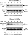
Ubiquitination does not affect pMHC-II endocytosis. Immature or in vitro-activated APCs were reversibly biotinylated on ice, and pMHC-II endocytosis was assayed as described in Materials and Methods. (A) Biotinylated immature BMDCs were cultured on ice (t = 0) or at 37 °C for various times. The cells were incubated in the absence or presence of glutathione on ice to remove surface-exposed biotin. Cells were lysed, and pMHC-II was isolated by immunoprecipitation and analyzed by SDS/PAGE and blotting for either biotin-labeled pMHC-II or total MHC-II. Only a fraction of the pMHC-II immunoprecipitate from cells incubated in the absence of glutathione on ice (t = 0) was loaded on the gel to assist in quantitative analysis of the blots. (B and C) The kinetics of pMHC-II endocytosis in immature (triangles) and in vitro-matured (squares) BMDCs (B) or resting (triangles) or in vitro-activated (squares) spleen B cells (C) was determined. The data shown are the mean ± SD obtained from three independent experiments. (D and E) The amount of surface pMHC-II internalized after a 15-min chase at 37 °C in wild-type or MHC-II K225R ubiquitination mutant immature BMDCs (D) or spleen B cells (E) was quantitated using our endocytosis assay. The data shown are the mean ± SD obtained from three independent experiments.
Fig. S1.
Fig. S1.
Ubiquitination does not affect pMHC-II endocytosis. The kinetics of pMHC-II endocytosis in immature and in vitro-matured BMDCs (A) or resting and in vitro-activated spleen B cells (B) was determined. Immature or in vitro-activated APCs were reversibly biotinylated on ice, and pMHC-II endocytosis was assayed as described in Materials and Methods. Biotinylated cells were cultured on ice (t = 0) or at 37 °C for various times. The cells were then incubated in the absence or presence of glutathione on ice to remove surface-exposed biotin. Cells were lysed, and pMHC-II was isolated by immunoprecipitation and analyzed by SDS/PAGE and blotting for either biotin-labeled pMHC-II or total MHC-II present in the immunoprecipitate. Only a fraction of the pMHC-II immunoprecipitate from cells incubated in the absence of glutathione on ice (t = 0) was loaded on the gels to assist in quantitative analysis of the blots. A representative blot from each condition is shown.
Fig. 3.
Fig. 3.
APC activation stimulates pMHC-II recycling in DCs and B cells. (A) Immature or LPS-matured BMDCs were reversibly biotinylated on ice, and pMHC-II recycling was assayed as described in Materials and Methods. BMDCs possessing internalized biotinylated pMHC-II were cultured on ice (t = 0) or at 37 °C for various times. The cells were placed on ice and incubated in the absence or presence of glutathione to remove surface-exposed biotin. Cells were lysed, and pMHC-II was isolated by immunoprecipitation and analyzed by SDS/PAGE and blotting for either biotin-labeled pMHC-II or total MHC-II. The data shown are the mean ± SD obtained from three independent experiments; *P < 0.05. (B) The reappearance of pMHC-II internalized in immature (I) and LPS-matured (M) BMDCs, freshly isolated (immature, I) and in vivo-activated (mature, M) spleen DCs, or resting (R) and in vitro-activated (A) spleen B cells after a 15-min chase period at 37 °C was quantitated using our recycling assay. In each panel, the data shown are the mean ± SD obtained from three independent experiments; *P < 0.05.
Fig. S2.
Fig. S2.
Activation of DCs stimulates pMHC-II recycling. Immature BMDCs (Upper) or LPS-matured BMDCs (Lower) were reversibly biotinylated on ice, and pMHC-II recycling was assayed as described in Materials and Methods. BMDCs possessing internalized biotinylated pMHC-II were cultured on ice (t = 0) or at 37 °C for various times. The cells were then placed on ice and incubated in the absence or presence of glutathione on ice to remove surface-exposed biotin. Cells were lysed, and pMHC-II was isolated by immunoprecipitation and analyzed by SDS/PAGE and blotting for either biotin-labeled pMHC-II or total MHC-II present in the immunoprecipitate. A representative blot from each condition is shown.
Fig. 4.
Fig. 4.
Ubiquitination regulates pMHC-II recycling in DCs and B cells. The extent of reappearance of internalized pMHC-II after a 15-min chase at 37 °C in immature BMDCs (A), freshly isolated spleen DCs (B), or freshly isolated spleen B cells (C) from the indicated mice was quantitated using our recycling assay. In each panel, the data shown are the mean ± SD obtained from three independent experiments; *P < 0.05, **P < 0.01.
Fig. S3.
Fig. S3.
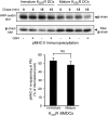
Activation-enhanced recycling is pMHC-II ubiquitination-dependent. Immature or LPS-matured BMDCs from MHC-II K225R ubiquitination mutant mice were reversibly biotinylated on ice, and pMHC-II recycling was assayed as described in Materials and Methods. Immature or LPS-matured BMDCs possessing internalized biotinylated pMHC-II were cultured on ice (t = 0) or at 37 °C for 15 min. The cells were then placed on ice and were incubated in the absence or presence of glutathione to remove surface-exposed biotin. Cells were lysed, and pMHC-II was isolated by immunoprecipitation and analyzed by SDS/PAGE and blotting for either biotin-labeled pMHC-II or total MHC-II present in the immunoprecipitate. (Upper) A representative blot from each condition is shown. (Lower) The extent of reappearance of internalized pMHC-II after a 15-min chase at 37 °C was quantitated using our recycling assay. The data shown are the mean ± SD obtained from three independent experiments; NS, not significant.
Fig. 5.
Fig. 5.
Ubiquitination promotes degradation of newly synthesized pMHC-II in DCs. (A) Immature BMDCs were pulse-labeled with [35S] for 1 h, washed, and chased in complete medium. After 2 h, either PBS or LPS was added to the cultures for an additional 18 h of chase at 37 °C. The recovery of [35S]-labeled pMHC-II was determined by immunoprecipitation, SDS/PAGE, and autoradiography. The abundance of [35S]–MHC-II β-chain present in each sample was quantitated by laser densitometry. The data shown are the mean ± SD obtained from three independent experiments; *P < 0.05. (B and C) Immature BMDCs isolated from wild-type and MHC-II K225R ubiquitination mutant mice (B) or wild-type and March-I KO mice (C) were pulse-labeled with [35S] for 1 h, washed, and chased for the indicated amount of time. The amount of [35S]–MHC-II β-chain present in each sample was determined as described above. The data shown are the mean ± SD obtained from three independent experiments; *P < 0.05.
Fig. 6.
Fig. 6.
Efficient recycling prolongs antigen presentation by DCs. Immature BMDCs obtained from either wild-type or MHC-II K225R ubiquitination mutant mice were treated with OVA323–339 peptide for 30 min on ice, washed, and then chased in complete medium at 37 °C for various times. DCs were harvested and cocultured with OVA-specific OT-II CD4 T cells at a 1:3 ratio. (A) After incubation of DCs and T cells for 18 h, cells were stained with CD69 and CD4 mAb, and the percentage of CD4 cells expressing high levels of surface CD69 was determined. (B) The relative extent of T-cell proliferation at each chase point was expressed as a fraction of that obtained by incubating the indicated DC type with OVA323–339 peptide-pulsed DCs. The data shown are the mean ± SD obtained from three independent experiments; **P < 0.01, ***P < 0.001.

References

    1. Trombetta ES, Mellman I. Cell biology of antigen processing in vitro and in vivo. Annu Rev Immunol. 2005;23:975–1028. - PubMed
    1. Banchereau J, et al. Immunobiology of dendritic cells. Annu Rev Immunol. 2000;18:767–811. - PubMed
    1. Roche PA, Furuta K. The ins and outs of MHC class II-mediated antigen processing and presentation. Nat Rev Immunol. 2015;15(4):203–216. - PMC - PubMed
    1. Rocha N, Neefjes J. MHC class II molecules on the move for successful antigen presentation. EMBO J. 2008;27(1):1–5. - PMC - PubMed
    1. Grant BD, Donaldson JG. Pathways and mechanisms of endocytic recycling. Nat Rev Mol Cell Biol. 2009;10(9):597–608. - PMC - PubMed

Publication types

LinkOut - more resources