. 2015 Aug 18;112(33):10389-94.
doi: 10.1073/pnas.1504625112. Epub 2015 Aug 3.

Nucleotides regulate the mechanical hierarchy between subdomains of the nucleotide binding domain of the Hsp70 chaperone DnaK

Affiliations

Nucleotides regulate the mechanical hierarchy between subdomains of the nucleotide binding domain of the Hsp70 chaperone DnaK

Daniela Bauer et al. Proc Natl Acad Sci U S A. .

Abstract

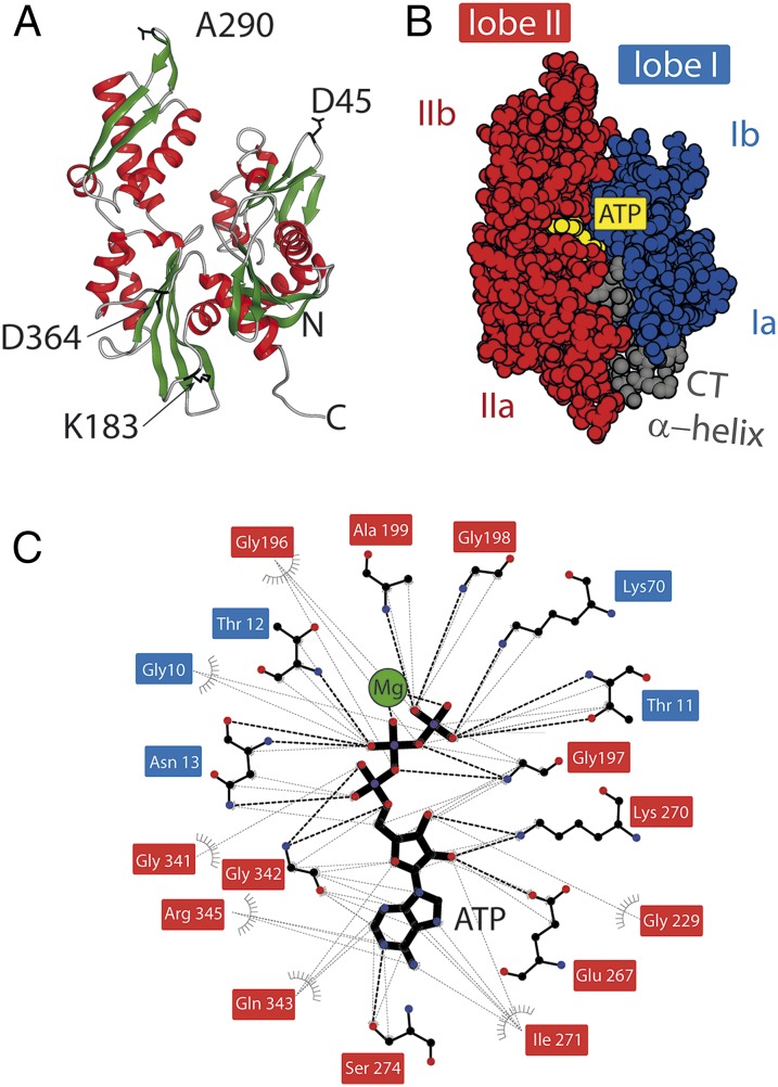
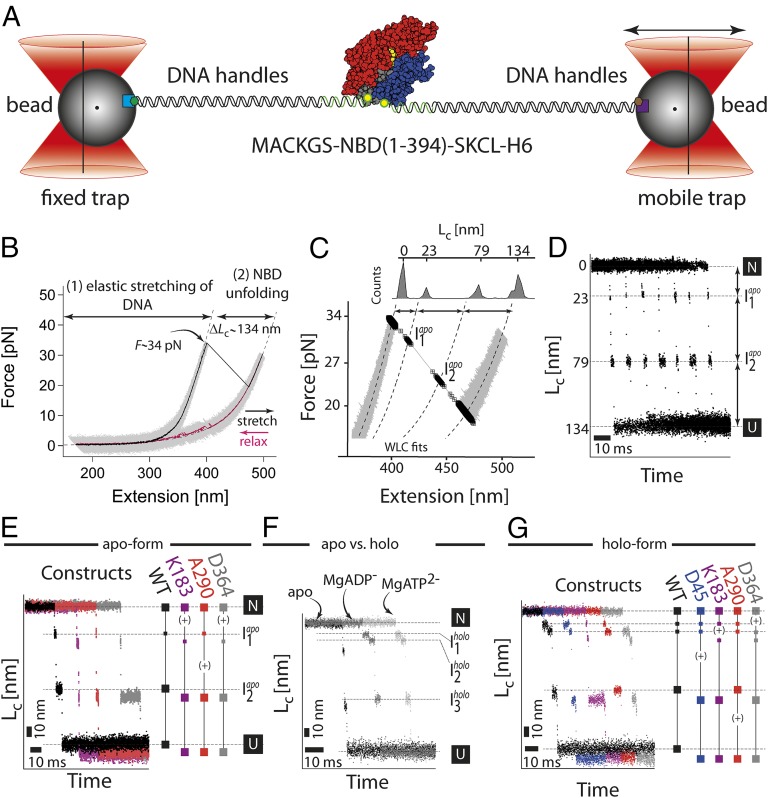
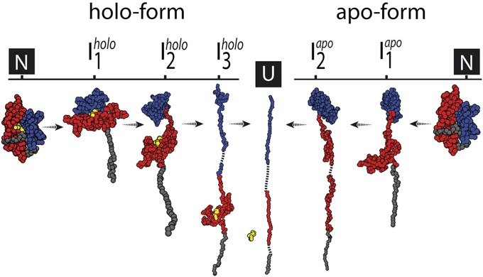
The regulation of protein function through ligand-induced conformational changes is crucial for many signal transduction processes. The binding of a ligand alters the delicate energy balance within the protein structure, eventually leading to such conformational changes. In this study, we elucidate the energetic and mechanical changes within the subdomains of the nucleotide binding domain (NBD) of the heat shock protein of 70 kDa (Hsp70) chaperone DnaK upon nucleotide binding. In an integrated approach using single molecule optical tweezer experiments, loop insertions, and steered coarse-grained molecular simulations, we find that the C-terminal helix of the NBD is the major determinant of mechanical stability, acting as a glue between the two lobes. After helix unraveling, the relative stability of the two separated lobes is regulated by ATP/ADP binding. We find that the nucleotide stays strongly bound to lobe II, thus reversing the mechanical hierarchy between the two lobes. Our results offer general insights into the nucleotide-induced signal transduction within members of the actin/sugar kinase superfamily.

Keywords: ATPase; elasticity; force; laser trapping; protein extension.


Conflict of interest statement

The authors declare no conflict of interest.

Figures

Fig. 1.
Fig. 1.
Structure of the NBD and the nucleotide binding pocket. (A) The 3D structure of the NBD from DnaK (PDB ID code 2KHO) (11). The position for the insertions (D45, K183, A290, and D364) are shown in black as a stick representation. (B) The protein comprises two lobes—lobe I (blue surface) and lobe II (red surface)—which are further subdivided into “a” and “b” subdomains. The lobes are connected by the C-terminal helix (gray surface). The ATP molecule (yellow spheres) binds between the lobes (PDB ID code 4B9Q) (39). The figures were prepared using DS Viewer (Accelrys Software, Inc.). (C) The analysis and mapping of the contacts between the protein’s residues and Mg2+ATP4- ligand (ball-and-stick representation, Mg2+ ion in green) using LigPlot+ software (42). The residue names have background-shading based on their correspondence to the lobes. Black broken lines indicate the hydrogen bonding between atoms; gray broken lines indicate hydrophobic/van der Waals contact.
Fig. 2.
Fig. 2.
Single molecule force experiments of the NBD by optical tweezers. (A) Optical trap assay. The protein is tethered to the beads by two DNA handles containing a corresponding epitope (biotin, green hexagon; digoxygenin, brown hexagon). The connection between the DNA and protein is realized by the modification of the two cysteine residues of the protein by the single-stranded DNA–maleimide oligonucleotide complementary to the DNA handle overhang. The 1 μm-sized functionalized (α-digoxygenin, purple square; streptavidin, light blue square) glass beads are trapped in the highly focused laser beam. One of the beams is reflected by the steerable mirror, which enables pulling and stretching of a single protein. (B) Force–extension curves of a single NBD domain (the apo-form). The protein–DNA construct was stretched (black curve, 1 kHz filtered) and relaxed (purple curve, 1 kHz filtered) at a constant velocity of 20 nm/s. The trace depicts two parts corresponding to the stretching of the DNA handles and a sudden rip at 34 pN corresponding to the unfolding of the protein. The dashed lines correspond to a WLC fit to the data yielding the contour-length increase of 134 nm. (C) A magnification of the force–extension trace. The unfolding phase of the protein reveals transient populations of the intermediates (black squares; for details, see Single Molecule Force Experiments on the NBD of the DnaK Chaperone). (D) Contour-length transformation of the force–extension data. Shown are multiple unfolding phases. (E) Contour-length increase versus time for the wild-type (black) and insert variants (purple for K183-Insert, red for A290-Insert, and gray for D364-Insert). The insert variants have an additional increase in the contour length (∼7 nm) due to insertion of a highly flexible 20 aa large loop. The plus sign (+) indicates an increase in the length at a particular position. (F) Comparison of the unfolding phases between different protein forms: apo, MgATP, and MgADP. The conditions were 50 mM Tris⋅HCl, 150 mM KCl, 5 mM MgCl2, and 1 mM ATP/ADP, pH 7.8. (G) Contour-length increase versus time for the wild-type (black) and insert variants (purple for K183-Insert, red for A290-Insert, and gray for D364-Insert) under holo-conditions (1 mM ATP, 5 mM MgCl2).
Fig. 3.
Fig. 3.
Forced unfolding simulations of the NBD. Simulation results for apo- and holo-NBD. (A) Force extension curve (FEC) for the unfolding of the apo-form. Intermediates Iapo1 and Iapo2 are labeled on the respective force peaks. (B) Tension propagation in the chain and conformational snapshots for the two intermediates evaluated from the red points in A. (Top) For Iapo1, tension is greatest in the connecting loop and in helices 15 and 16 (residues 168–187 and 339–383). The snapshot shows that this corresponds to the separation of the lobes and the unfolding of those helices. (Bottom) For Iapo2, before lobe I unravels, the connecting loop and the entire lobe II experiences increased tension (residues from Iapo1 plus 188–338). (C) FEC for the unfolding of the holo-form. Intermediates Iholo1, Iholo2, and Iholo3 are labeled. (D) Tension propagation in the chain and conformational snapshots for the three intermediates evaluated from the red points in C. (Top) For Iholo1, helices 15 and 16 are extracted from lobe II due to increased tension (residues 346–383). (Middle) For Iholo2, the two lobes separate due to the increased tension in the connecting loop (residues from Iholo1 plus 169–187). (Bottom) In Iholo3, only lobe II remains folded (residues from Iholo2 and positions 1–168 unfold).
Fig. 4.
Fig. 4.
Summary of the unfolding pathway of the NBD of DnaK. The structures of intermediates are those from the pulling simulations. The red surface indicates lobe II; the blue surface indicates lobe I and the yellow sphere is an ADP molecule.

References

    1. Smock RG, Gierasch LM. Sending signals dynamically. Science. 2009;324(5924):198–203. - PMC - PubMed
    1. Janovjak H, Sapra KT, Kedrov A, Muller DJ. From valleys to ridges: Exploring the dynamic energy landscape of single membrane proteins. Chemphyschem. 2008;9(7):954–966. - PubMed
    1. Kedrov A, Ziegler C, Muller DJ. Differentiating ligand and inhibitor interactions of a single antiporter. J Mol Biol. 2006;362(5):925–932. - PubMed
    1. Kedrov A, Krieg M, Ziegler C, Kuhlbrandt W, Muller DJ. Locating ligand binding and activation of a single antiporter. EMBO Rep. 2005;6(7):668–674. - PMC - PubMed
    1. Manibog K, Li H, Rakshit S, Sivasankar S. Resolving the molecular mechanism of cadherin catch bond formation. Nat Commun. 2014;5:3941. - PubMed

Publication types

MeSH terms

LinkOut - more resources